PF3D7_1218000 thrombospondin-related apical membrane protein (TRAMP)
Disruptability [+]
| Species | Disruptability | Reference | Submitter | |
|---|---|---|---|---|
| P. falciparum 3D7 |
Refractory |
USF piggyBac screen (Insert. mut.) | USF PiggyBac Screen | |
| P. berghei ANKA |
Refractory |
RMgm-805 | Imported from RMgmDB | |
| P. berghei ANKA |
Refractory |
RMgm-505 | Imported from RMgmDB | |
| P. berghei ANKA |
Refractory |
PlasmoGEM (Barseq) | PlasmoGEM | |
Mutant phenotypes [+]
| Species | Stage | Phenotype | Reference | Submitter |
|---|---|---|---|---|
| P. falciparum 3D7 | Asexual |
Invasion defect |
36396942 (Conditional)
\"Analysis of parasite growth showed they were not able to expand, indicating the function of each protein was essential (Fig. 1d–f and Extended Data Fig. 1g–i) and that the schizont to ring stage transition was blocked, consistent with these proteins being required for invasion (Fig. (Fig.2a2a).\" |
Theo Sanderson, Francis Crick Institute |
Imaging data (from Malaria Metabolic Pathways)
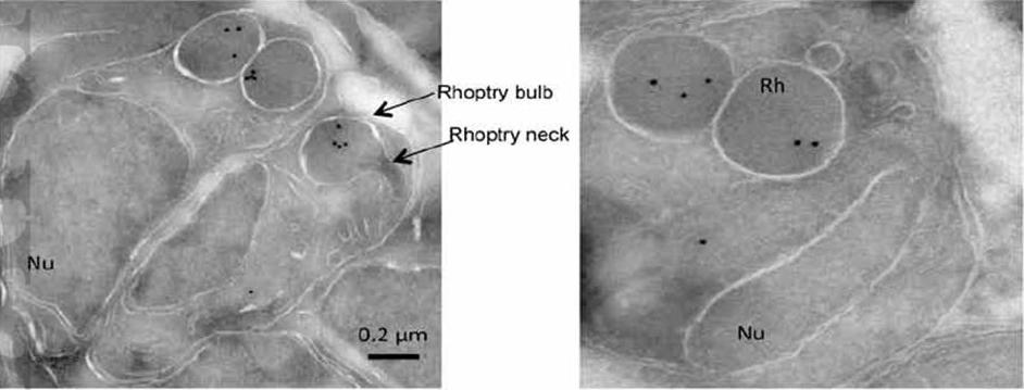
Localization of PfTRAMP by immuno-electron microscopy. Electron micrographs showing immuno-gold staining of P. falciparum schizonts (A) and merozoites (B) using anti- PfTRAMP antibodies. PfTRAMP localizes to the bulb region of rhoptries in merozoites developing within a schizont (A) as well as free merozoites (B).(Rh: rhoptry, Nu: nucleus).Siddiqui FA, Dhawan S, Singh S, Singh B, Gupta P, Pandey A, Mohmmed A, Gaur D, Chitnis CE. A Thrombospondin Structural Repeat Containing Rhoptry Protein from Plasmodium falciparum Mediates Erythrocyte Invasion. Cell Microbiol. 2013 15(8):1341-56
See original on MMP
PfTRAMP localizes to the rhoptries of P. falciparum schizonts and merozoites.(A) Co-localization of PfTRAMP with MSP1, EBA175 and Rh2b in schizonts and free merozoites by IFA. PfTRAMP (green) co-localizes with rhoptry marker PfRh2b (red) but does not co-localize with EBA175 (red) or merozoite surface marker MSP1 (red) in schizonts and merozoites. (B)Co-localization of PfTRAMP-GFP in transgenic P. falciparum 3D7 with MSP1, AMA1 and Clag3.1. PfTRAMP-GFP (green) co-localizes with rhoptry marker Clag 3.1 (red) but not with microneme markerPfAMA1 (red) or merozoite suface marker PfMSP1 (red) in schizonts and merozoites. Nuclei were stained with DNA intercalating dye DAPI.Siddiqui FA, Dhawan S, Singh S, Singh B, Gupta P, Pandey A, Mohmmed A, Gaur D, Chitnis CE. A Thrombospondin Structural Repeat Containing Rhoptry Protein from Plasmodium falciparum Mediates Erythrocyte Invasion. Cell Microbiol. 2013 15(8):1341-56
See original on MMP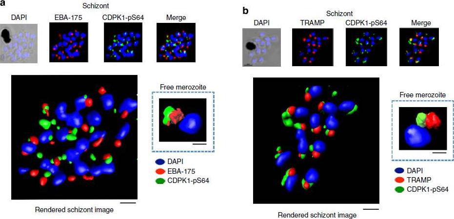
PfCDPK1 phosphorylated at serine-64 is associated with apical parasite structures. (a) A schizont stage parasite stained with DAPI to reveal the nuclei (blue), an antibody to PfEBA-175 to reveal the micronemes (red) and the phospho-specific CDPK1-pS64 antibody (green). A merge of images from all three stains is shown on the far right; this is a representative image from at least three experiments. Also shown is a rendered image where deconvoluted z stacks were reconstructed in 3D, with interpolation. The inset shows a rendered image of a free merozoite from the same preparation . (b) The same as a, but instead of probing with an anti-EBA-175 antibody the preparation was probed with an antibody to the rhoptrymarker PfTRAMP . Scale bars, a-1 mm (insert-0.5 mm), b-1 mm (insert-0.5 mm).Alam MM, Solyakov L, Bottrill AR, Flueck C, Siddiqui FA, Singh S, Mistry S, Viskaduraki M, Lee K, Hopp CS, Chitnis CE, Doerig C, Moon RW, Green JL, Holder AA, Baker DA, Tobin AB. Phosphoproteomics reveals malaria parasite Protein Kinase G as a signalling hub regulating egress and invasion. Nat Commun. 2015 6:7285
See original on MMP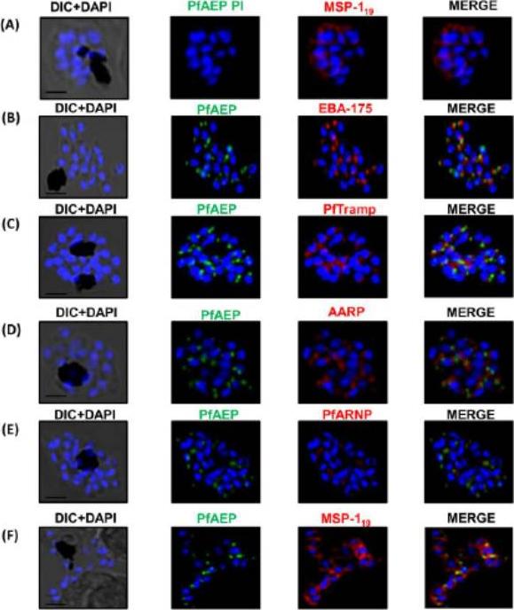
PfAEP localization in schizont stages analyzed by confocal microscopy. Subcellular localization of PfAEP was studied by co-staining with antibodies against micronemal protein EBA-175 (B), rhoptry bulb protein PTRAMP (C), and rhoptry neck,apical asparagine rich protein (AARP) (D), apical rhoptry neck protein (ARNP) (E), and merozoite surface protein (MSP) (F).As a control, pre-immune serum of PfAEP was checked and no staining was observed in schizonts (A). Mature schizonts were costained with anti-PfAEP (green) and anti-EBA-175, TRAMP, AARP, ARNP, MSP-119 antibodies (red). The nuclei of schizonts were stained with DAPI (blue) and visualized by Nikon N-SIM confocal microscope. All apical marker proteins and PfAEP showed punctate staining in schizonts distinct from DAPI. Co-staining of PfAEP with surface marker MSP-119 showed PfARNP localized at the apical tip with surface of merozoites stained by MSP-119. PfAEP staining did not merge with either markers of microneme or rhoptry indicating that PfAEP is neither a resident of micronemes or rhoptry ofmerozoites. Scale bar 2 mm.Hans N, Relan U, Dubey N, Gaur D, Chauhan VS. Identification and localization of a Novel Invasin of Plasmodium falciparum. Mol Biochem Parasitol. 2015 Sep 29. [Epub ahead of print]
See original on MMP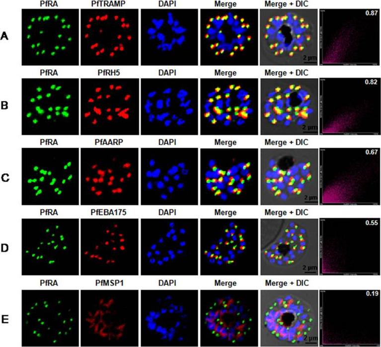
PfRA localizes to the rhoptries of P. falciparum schizonts and merozoites. Co-localization of PfRA with (A) PfTRAMP, (B) PfRH5, (C) PfAARP, (D) PfEBA-175 and (E) MSP-1 in schizont stage parasites by confocal immunoflourescence microscopy. PfRA (green) co-localizes with rhoptry bulb protein PfTRAMP(red) and PfRH5 (red) but does not co-localize with the micronemal protein PfEBA-175 (red) and merozoite surface protein MSP-1 in schizonts. PfRA (green) showed partial co-localization with rhoptry neck protein PfAARP (red). The co-localization of PfRA was quantitatively analyzed and the Pearson correlation co-efficientwith PfTRAMP (0.87) and PfRH5 (0.82) suggested that the two proteins were co-localized with PfRA. The coefficients of PfRA with PfAARP, PfEBA-175 and MSP1 were lower than 0.7 indicating that the three proteins were not co-localized with PfRA. Nuclei were stained with DNA intercalating dye DAPI. (Scale bar, 2 μm.)Anand G, Reddy KS, Pandey AK, Mian SY, Singh H, Mittal SA, Amlabu E, Bassat Q, Mayor A, Chauhan VS, Gaur D. A novel Plasmodium falciparum rhoptry associated adhesin mediates erythrocyte invasion through the sialic-acid dependent pathway. Sci Rep. 2016 6:29185.
See original on MMPMore information
| PlasmoDB | PF3D7_1218000 |
| GeneDB | PF3D7_1218000 |
| Malaria Metabolic Pathways | Localisation images Pathways mapped to |
| Previous ID(s) | 2277.t00174, MAL12P1.174, PFL0870w |
| Orthologs | PBANKA_1433600 , PCHAS_1435600 , PKNH_1437600 , PVP01_1436800 , PVX_123575 , PY17X_1436000 |
| Google Scholar | Search for all mentions of this gene |