PF3D7_1215000 thioredoxin peroxidase 2 (Trx-Px2)
Disruptability [+]
| Species | Disruptability | Reference | Submitter | |
|---|---|---|---|---|
| P. falciparum 3D7 |
Refractory |
USF piggyBac screen (Insert. mut.) | USF PiggyBac Screen | |
| P. berghei ANKA |
Possible |
RMgm-791 | Imported from RMgmDB | |
| P. berghei ANKA |
Possible |
PlasmoGEM (Barseq) | PlasmoGEM | |
Mutant phenotypes [+]
| Species | Stage | Phenotype | Reference | Submitter |
|---|---|---|---|---|
| P. berghei ANKA | Asexual |
No difference |
RMgm-791 | Imported from RMgmDB |
| P. berghei ANKA | Asexual |
No difference |
PlasmoGEM (Barseq) | PlasmoGEM |
| P. berghei ANKA | Gametocyte |
No difference |
RMgm-791 | Imported from RMgmDB |
| P. berghei ANKA | Oocyst |
No difference |
RMgm-791 | Imported from RMgmDB |
Imaging data (from Malaria Metabolic Pathways)
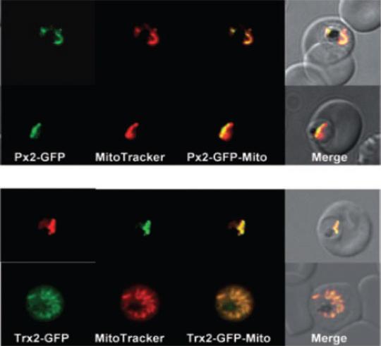
Localization of PfTrx-Px2 and PfTrx2 in P. falciparum erythrocytic stages. P. falciparum erythrocytic stages were transfected with construct pHH2-Px2-GFP, pHH2-Trx2-GFP leading to the expression of the peroxiredoxin or thioredoxin2 C-terminally fused to green fluorescent protein (GFP).A. The localization of the peroxiredoxin-GFP fusion protein in parasites previously treated with MitoTracker CMX-Ros was analysed by fluorescence light microscopy. Phase, phase contrast of parasitized erythrocytes infected with P. falciparum trophozoites; PfTrx-Px2-GFP, parasitized erythrocytes expressing the peroxiredoxin-GFP fusion protein analysed using the FITC channel; MitoTracker, parasitized erythrocytes expressing the peroxiredoxin-GFP fusion protein analysed using the rhodamine channel; PfTrx-Px2-Mito; merge of FITC and rhodamine channels; merge, merge of all images. The images show that the peroxiredoxin-GFP fusion protein is colocalizing with the mitochondrion (stained by MitoTracker).B. The expression of pHH2-PfTrx2 results in the localization of the fusion protein the mitochondrion. Phase, phase contrast of parasitized erythrocytes infected with P. falciparum trophozoites; PfTrx2-GFP, parasitized erythrocytes expressing the Trx2-GFP fusion protein analysed using the FITC channel; MitoTracker, parasitized erythrocytes expressing the Trx2-GFP fusion protein analysed using the rhodamine channel; PfTrx2-Mito; merge of FITC and rhodamine channels; merge, merge of all images.Boucher IW, McMillan PJ, Gabrielsen M, Akerman SE, Brannigan JA, Schnick C, Brzozowski AM, Wilkinson AJ, Müller S. Structural and biochemical characterization of a mitochondrial peroxiredoxin from Plasmodium falciparum. Mol Microbiol. 2006 61:948-59.PMID
See original on MMP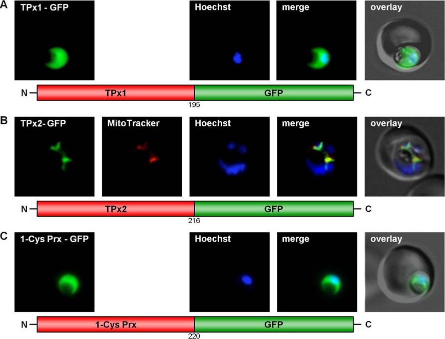
GFP targeting by various P. falciparum peroxiredoxins. (A) Cytosolic localization of TPx1. (B) Mitochondrial targeting of TPx2. (C) Cytosolic localization of 1-Cys Prx. Live cell imaging of erythrocytes infected with transgenic parasites for solely cytosolic GFP signals. Colocalization of GFP with the mitochondrial stain MitoTrackerOrange in fixed cells.Kehr S, Sturm N, Rahlfs S, Przyborski JM, Becker K. Compartmentation of redox metabolism in malaria parasites. PLoS Pathog. 2010 6:e1001242.
See original on MMP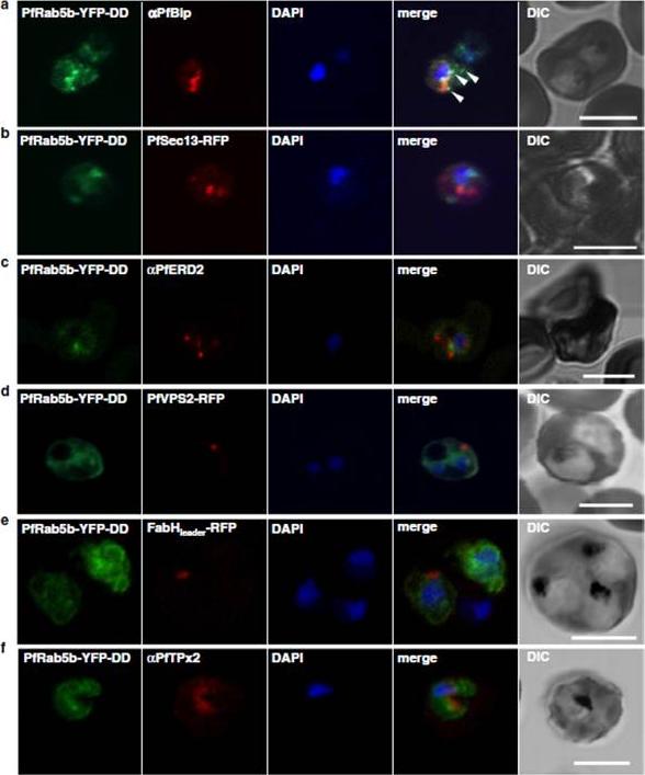
Localization of PfRab5b to a punctate compartment in the parasite cytoplasm. Triple staining with PfRab5b-YFP-DD (green), DAPI (blue) and one of the following markers (red): PfBip (a, ER), PfSec13-RFP (b, ER exit site), PfERD2 (c, Golgi), PfVPS2-RFP (d, putative multivesicular body/endosome), FabHleader-RFP (e, apicoplast), or PfTPx-2 (f, mitochondria) after 24 h incubation with Shld1. PfRab5b-YFP-DD localized adjacent to the Bip signal (arrowheads). Bars 5 μm.Ebine K, Hirai M, Sakaguchi M, Yahata K, Kaneko O, Saito-Nakano Y. Plasmodium Rab5b is secreted to the cytoplasmic face of the tubovesicular network in infected red blood cells together with N-acylated adenylate kinase 2. Malar J. 2016 15:323.
See original on MMPMore information
| PlasmoDB | PF3D7_1215000 |
| GeneDB | PF3D7_1215000 |
| Malaria Metabolic Pathways | Localisation images Pathways mapped to |
| Previous ID(s) | 2277.t00146, MAL12P1.145, PFL0725w |
| Orthologs | PBANKA_1430800 , PCHAS_1432700 , PVP01_1433800 , PVX_123435 , PY17X_1433000 |
| Google Scholar | Search for all mentions of this gene |