PF3D7_1203700 nucleosome assembly protein (NAPL)
Disruptability [+]
| Species | Disruptability | Reference | Submitter | |
|---|---|---|---|---|
| P. falciparum 3D7 |
Refractory |
19176479 | Theo Sanderson, Wellcome Trust Sanger Institute | |
| P. falciparum 3D7 |
Refractory |
USF piggyBac screen (Insert. mut.) | USF PiggyBac Screen | |
| P. berghei ANKA |
Refractory |
PlasmoGEM (Barseq) | PlasmoGEM | |
Mutant phenotypes [+]
None reported yet. Please press the '+' button above to add one.Imaging data (from Malaria Metabolic Pathways)
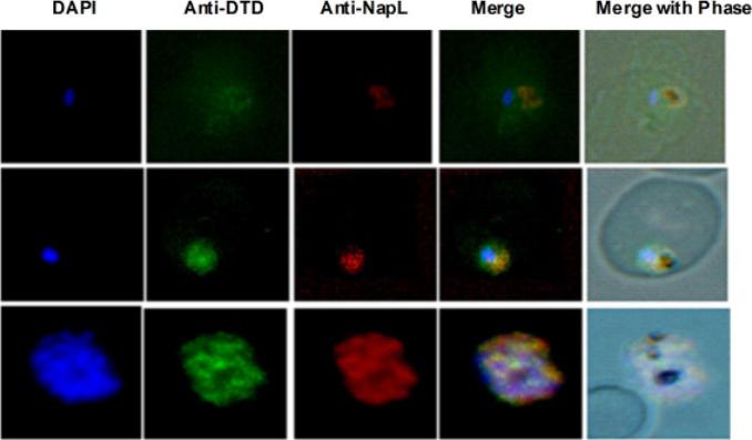
Expression and activity of PfDTD. Localization of PfDTD in different intra-erythrocytic stages of P. falciparum by immunofluo-rescence staining, Ring stage (R), trophozoite stage (T), and schizont stage (S) are shown. In each panel are shown: (i) image of cell stained with DAPI (blue), (ii) anti-PfDTD antibodies, (iii) anti-PfNapL antibodies, (iv) merged image, (v) merge with phase contrast. Results clearly show the colocalization of PfDTD with PfNapL at all the asexual life stages of parasite, suggesting PfDTD to be cytoplasmic in nature, like for PfNapL.Bhatt TK, Yogavel M, Wydau S, Berwal R, Sharma A. Ligand-bound structures provide atomic snapshots for the catalytic mechanism of D-amino acid deacylase. J Biol Chem. 2010 Feb 285(8):5917-30.
See original on MMP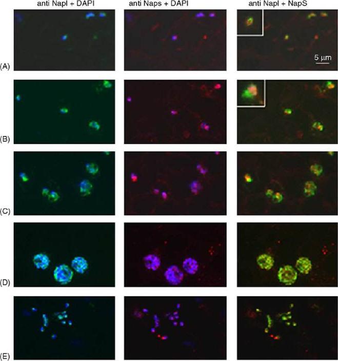
Double immunofluorescence analysis of PfNAPL and PfNAPS in asexual and sexual parasites. Acetone fixed parasites were reacted with rabbit anti-NAPL antibodies and a rat anti-NAPS antibody (1:400). Anti-PfNAPL fluorescence (green, first column) and anti-PfNAPS fluorescence (red, second column) were obtained reacting the samples with fluorescein-conjugated anti rabbit IgG antiserum and rhodamine-conjugated anti rat IgG antiserum (1:200) (Cappel), and are combined with DAPI fluorescent signal. Composite picture of PfNAPL and PfNAPS fluorescent signals is shown in the “merge” column, in which DAPI fluorescence is not shown for clarity. Panel A: early rings; B: late rings; C: trophozoites/early schizonts; D: schizonts; E: free merozoites; Inserts at the right end in panels A and B contain higher magnification of an individual parasite at that time point. Bar is 5mm. PfNAPS did not co-localise with PfNAPL and was more intimately associated with the parasite nucleus,Chandra BR, Olivieri A, Silvestrini F, Alano P, Sharma A. Biochemical characterization of the two nucleosome assembly proteins from Plasmodium falciparum. Mol Biochem Parasitol. 2005 142:237-47. Copyright Elsevier 2009.
See original on MMP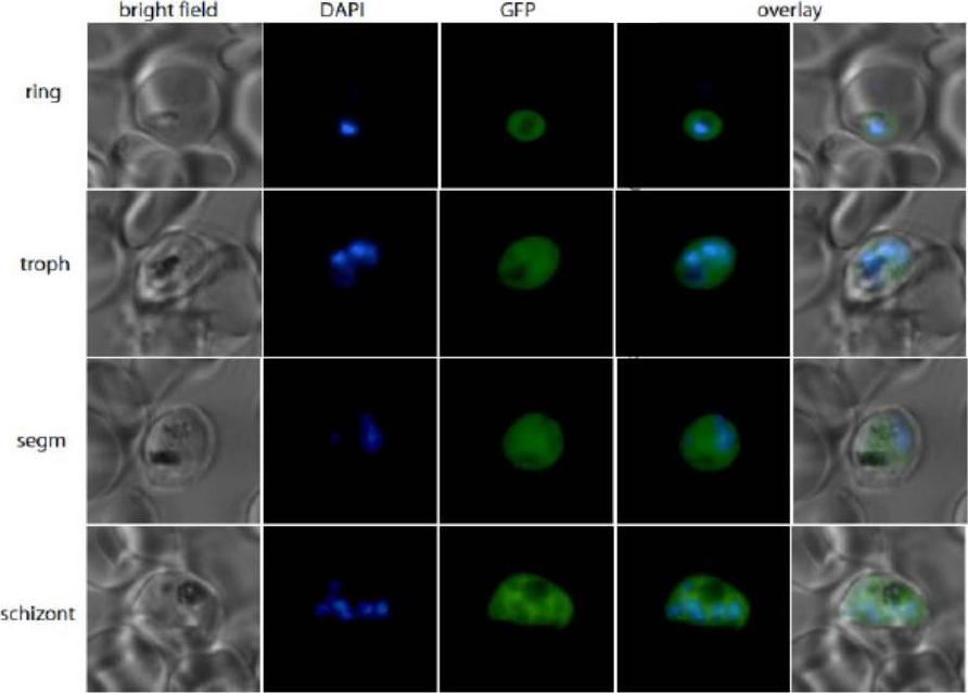
Stage specific expression of PfNapL-GFP chimeras. The localization of (b) PfNapL wild-type-GFP, (c) PfNapL L46A-GFP and (d) PfNapL Q50A in different stages of the erythrocytic life cycle of P. falciparum is shown. The first column of each panel shows bright field pictures, followed by a nuclear DAPI stain, GFP fluorescence, an overlay of the nuclear and GFP localization and - in the last column - overlay of bright field with DAPI and GFP. PfNapL wild-type-GFP was etected in the cytoplasm of the parasites.Gill J, Yogavel M, Kumar A, Belrhali H, Jain SK, Rug M, Brown M, Maier AG, Sharma A. Crystal structure of malaria parasite nucleosome assembly protein (NAP): distinct modes of protein localization and histone recognition. J Biol Chem. 2009 284:10076-87.
See original on MMP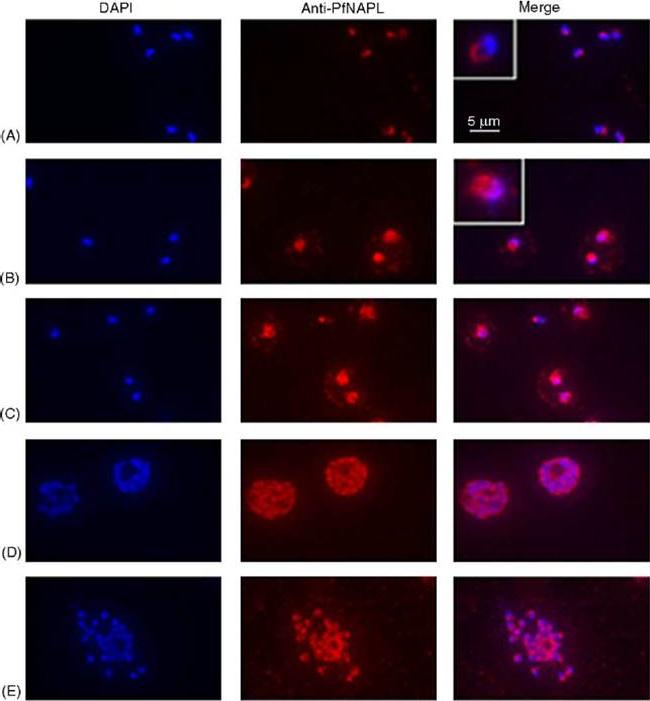
Immunofluorescence analysis of PfNAPLin asexual and sexual parasites. Acetone fixed parasites from a synchronous asexual culture reacted with rabbit anti-PfNAPL antibodies (1:400), rhodamine-conjugated anti rabbit IgG antiserum (1:200), and DAPI (0.1mgml−1). Fields A–E are photographed with filters specific for rhodamine and DAPI fluorescences, and combined fluorescent signals are shown in the “merge” column. A: early rings; B: late rings; C: trophozoites/early schizonts; D: schizonts; E: free merozoites. Inserts at the right end in panels A and B contain higher magnification of an individual parasite at that time point. Bar is 5 mm. PfNAPL was expressed in all erythrocytic stages of the parasite and predominantly localized in the cytoplasm in asexual and sexual stages of the parasite.Chandra BR, Olivieri A, Silvestrini F, Alano P, Sharma A. Biochemical characterization of the two nucleosome assembly proteins from Plasmodium falciparum. Mol Biochem Parasitol. 2005 142:237-47. Copyright Elsevier 2009.
See original on MMP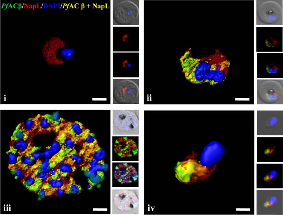
Expression of PfACb in P. falciparum blood stages. Immunofluorescence assays (IFA) were used to detect PfACb (green) in P. falciparum rings (i), trophozoites (ii), schizonts (iii) and merozoites (iv) using mouse antisera against a peptide derived from PfACb. Nuclear DNA was stained with DAPI (blue). Rabbit antiserum against P. falciparum cytoplasmic protein PfNAPL (red) was used for co-localization. Yellow indicates overlap of PfACb and PfNAPL. PfACb is thus expressed in P. falciparum merozoites and is localized in the cytoplasm.Dawn A, Singh S, More KR, Siddiqui FA, Pachikara N, Ramdani G, Langsley G, Chitnis CE. The Central Role of cAMP in Regulating Plasmodium falciparum Merozoite Invasion of Human Erythrocytes. PLoS Pathog. 2014 18;10(12):e1004520.
See original on MMP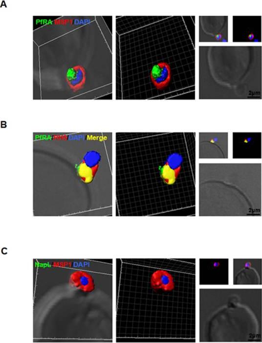
PfRA is present at the apical surface of an invading merozoite. Confocal immunofluorescencemicroscopy analysis of PfRA localization in cytochalasin-D treated invading merozoites under non-permeable fixing conditions using glutaraldehyde /paraformaldehyde. (A) 3D reconstruction of z-stacks during merozoite invasion co-immunostained with MSP-1and PfRA. PfRA detected at the apical end of the merozoite surface. In the 3D images, the γ settings were altered for visual representation only. (B) PfRA (green) co-localizes with rhoptry bulb marker PfRH5 (red) that is known to be translocated to the merozoite surface to engage with its erythrocyte receptor, Basigin. (C) As a control, staining of a nuclear parasite protein, NapL, was not detected under the same non-permeable fixing conditions confirming that the fluorescent signals were obtained only from surface localized proteins. Nuclei were stained with DNA intercalating dye DAPI (Scale bar, 2 μ m.)Anand G, Reddy KS, Pandey AK, Mian SY, Singh H, Mittal SA, Amlabu E, Bassat Q, Mayor A, Chauhan VS, Gaur D. A novel Plasmodium falciparum rhoptry associated adhesin mediates erythrocyte invasion through the sialic-acid dependent pathway. Sci Rep. 2016 6:29185.
See original on MMPMore information
| PlasmoDB | PF3D7_1203700 |
| GeneDB | PF3D7_1203700 |
| Malaria Metabolic Pathways | Localisation images Pathways mapped to |
| Previous ID(s) | 2277.t00037, MAL12P1.37, PFL0185c |
| Orthologs | PBANKA_0602700 , PCHAS_0604500 , PKNH_1303500 , PVP01_1303000 , PVX_084230 , PY17X_0605200 |
| Google Scholar | Search for all mentions of this gene |