PF3D7_1130200 60S ribosomal protein P0 (PfP0)
Disruptability [+]
| Species | Disruptability | Reference | Submitter |
|---|---|---|---|
| P. falciparum 3D7 |
Refractory |
USF piggyBac screen (Insert. mut.) | USF PiggyBac Screen |
Mutant phenotypes [+]
None reported yet. Please press the '+' button above to add one.Imaging data (from Malaria Metabolic Pathways)
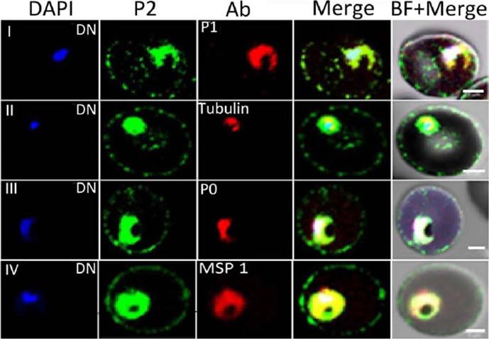
Immunofluorescence assay of P. falciparum infected erythrocytes (IE) using various antibodies. Localization of DAPI (blue), P2 (green); various antibodies (anti-PfP1, anti b-tubulin, anti-PfP0, and anti-MSP1) in red at the di-nuclear (DN) stage of Plasmodium falciparum infected erythrocytes. Scale bar indicates 2 mm.Das S, Basu H, Korde R, Tewari R, Sharma S. Arrest of Nuclear Division in Plasmodium through Blockage of Erythrocyte Surface Exposed Ribosomal Protein P2. PLoS Pathog. 2012 Aug;8(8):e1002858.
See original on MMP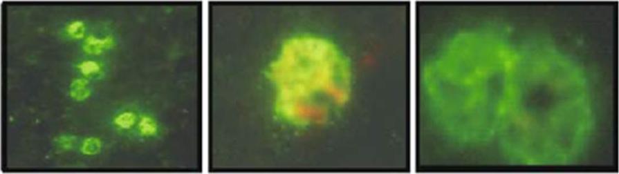
Localization of PfP0 in late schizonts as determined by indirect immunofluorescence assays. Purified final bleeding sera were used as primary antibody. PfP0 showed a grapelike fluorescence pattern typical of merozoite surface proteins.Arevalo-Pinzon G, Curtidor H, Reyes C, Pinto M, Vizcaíno C, Patarroyo MA, Patarroyo ME. Fine mapping of Plasmodium falciparum ribosomal phosphoprotein PfP0 revealed sequences with highly specific binding activity to human red blood cells. J Mol Med (Berl). 2010 88:61-74.
See original on MMP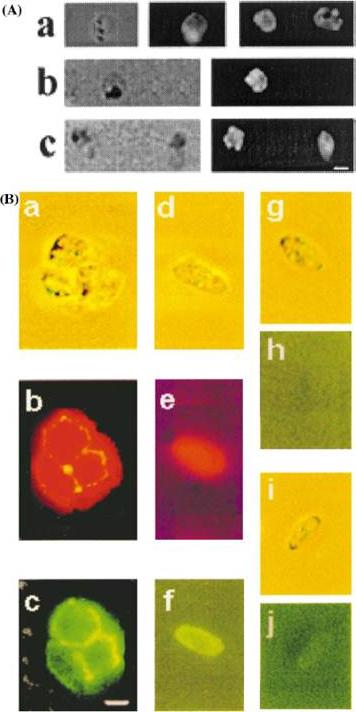
Immunofluorescence microscopy of P. falciparum merozoites and sexual stages. Upper panel A: fresh merozoite preparations were treated with anti-PfP0N (a, N-terminal), anti-PfP0C (b, C-terminal) and anti-MSP1 antibodies, each at a dilution of 1:50. Bound antibodies were visualized with anti-rabbit IgG-FITC secondary antibodies. The left hand panels show the bright field, and the right hand panels show the immunofluorescence. Bar corresponds to 1 mm. Lower Panel B: co-staining immunofluorescence analysis of macro gametes and red cell-free gametocytes. Panel a (bright field); b and c (corresponding immunofluorescent images) of a clump of two gametes and one gametocyte. Panel d (bright field), e and f (corresponding immunofluorescent images) of a single gametocyte. Panel g and i are the bright fields showing a gametocyte, while h andj are the corresponding immunofluorescent images, respectively.Panels a–f show the parasites treated with rabbit anti-PfP0N and Mab anti-Pfs230 [21]; g and h are gametocytes treated with rabbit pre-immune sera, and i and j are gametocytes treated with Mab anti-Pfg27, an internal marker of sexual stages . Anti-PfP0N was visualized with anti- rabbit IgG rhodamine secondary antibodies (b, e), while the monoclonal antibodies were visualized with anti-mouse IgG-FITC secondary antibodies (c, f and j). Rabbit pre-immune serum (panel h) was visualized with anti-rabbit IgG-FITC. All antibodies were used at 1:100 dilution. Bar corresponds to 5 mm.Chatterjee S, Singh S, Sohoni R, Kattige V, Deshpande C, Chiplunkar S, Kumar N, Sharma S. Characterization of domains of the phosphoriboprotein P0 of Plasmodium falciparum. Mol Biochem Parasitol. 2000 107(2):143-54.
See original on MMPMore information
| PlasmoDB | PF3D7_1130200 |
| GeneDB | PF3D7_1130200 |
| Malaria Metabolic Pathways | Localisation images Pathways mapped to |
| Previous ID(s) | PF11_0313 |
| Orthologs | PBANKA_0918000 , PCHAS_0928100 , PKNH_0928100 , PVP01_0930800 , PVX_092120 , PY17X_0919700 |
| Google Scholar | Search for all mentions of this gene |