PF3D7_1129100 parasitophorous vacuolar protein 1 (PV1)
Disruptability [+]
| Species | Disruptability | Reference | Submitter | |
|---|---|---|---|---|
| P. falciparum 3D7 |
Possible |
USF piggyBac screen (Insert. mut.) | USF PiggyBac Screen | |
| P. berghei ANKA |
Possible |
PlasmoGEM (Barseq) | PlasmoGEM | |
| P. berghei ANKA |
Possible |
RMgm-4189 | Imported from RMgmDB | |
| P. berghei ANKA |
Possible |
RMgm-4536 | Imported from RMgmDB | |
Mutant phenotypes [+]
| Species | Stage | Phenotype | Reference | Submitter |
|---|---|---|---|---|
| P. falciparum 3D7 | Asexual |
Altered cytoadherence |
https://www.nature.com/articles/ncomms16044
(Knock down)
Viable upon knockdown with no growth rate difference in vitro. But decreased binding to CD36, less deformability, and less protein export. |
Theo Sanderson, Wellcome Trust Sanger Institute |
| P. berghei ANKA | Asexual |
Attenuated |
PlasmoGEM (Barseq) | PlasmoGEM |
| P. berghei ANKA | Asexual |
Difference from wild-type |
RMgm-4189
Growth and multiplication of blood stages in mice comparable to wild type parasites. Two separate clones of mice infected with PbDPV1 parasites succumbed to cerebral malaria 24 days later than mice infected with wild-type PbANKA (PbANKA 57 days post infection, PbDPV1 79 days post infection). |
Imported from RMgmDB |
| P. berghei ANKA | Asexual |
No difference |
RMgm-4536 | Imported from RMgmDB |
| P. berghei ANKA | Oocyst |
No difference |
RMgm-4536 | Imported from RMgmDB |
Imaging data (from Malaria Metabolic Pathways)
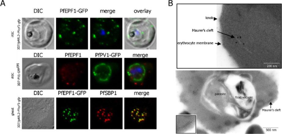
Localization and topology of the PfEPF1 proteins. A. Fluorescent patterns of iRBCs infected by 3D7/pARL2-Pfepf1-gfp (live imaging) and 3D7/PV1-GFPEPI (GFP fluorescence, in green, and immunodetection using anti-PfEPF1 antibodies, in red) and of resealed ghosts from 3D7/pARL2-Pfepf1-gfp iRBCs (GFP fluorescence, in green, and immunodetection using anti-SBP1, in red, antibodies). RBCs and ghost preparations were incubated with DAPI for nucleus labelling. B. Immunoelectron microscopy using anti-GFP antibodies. The inset shows higher magnification of a labelled Maurer’s cleft. Fluorescent dots in the iRBC cytoplasm; some at the erythrocyte periphery are suggestive of Maurer’s clefts while others are observed at the periphery of the parasitophorous vacuole. The Maurer’s cleft localization of the protein was confirmed by its colocalization with PfSBP1.Mbengue A, Audiger N, Vialla E, Dubremetz JF, Braun-Breton C. Novel Plasmodium falciparum Maurer's clefts protein families implicated in the release of infectious merozoites. Mol Microbiol. 2013 88(2):425-42
See original on MMP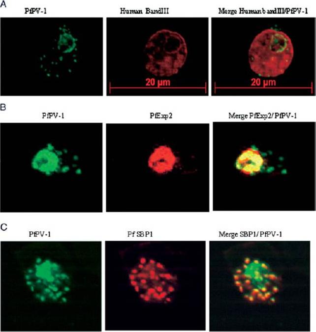
Localization of PfPV-1 in IRBCs. Trophozoite-IRBCs were spread onto microscope slides. Cells were fixed and incubated with mAbs that were directed either against the band 3 protein of the erythrocyte membrane (A), against the vacuolar marker protein PfEXP-2 (B), or against the Maurer’s clefts marker protein PfSBP1 (C). All slides were also incubated with the polyclonal antiserum against PfPV-1. Binding of the mAbs was monitored with Cy3 conjugated anti-mouse secondary antibodies (red), and binding of the polyclonal antiserum was monitored with Cy2 conjugated anti-rabbit secondary antibodies (green). PfPV-1 is concentrated in a distinct ring surrounding the parasite cell, and this location is distinct from that of the erythrocyte membrane (A). It colocalizes with the vacuolar marker protein PfEXP-2 (B) and with PfSBP1 (C), suggesting that it is present not only in the PV but also in Maurer’s clefts.Nyalwidhe J, Lingelbach K. Proteases and chaperones are the most abundant proteins in the parasitophorous vacuole of Plasmodium falciparum-infected erythrocytes. Proteomics. 2006 6:1563-73.
See original on MMP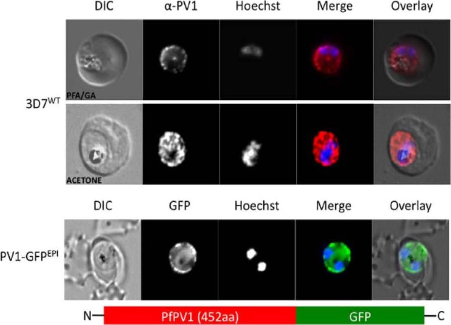
Localisation of PfPV1. (A, upper and middle panel) Immunofluorescence localisation using specific anti-PfPV1 antibodies. Lower panel: Episomal expression of a PfPV1-GFP chimera. DIC, differential interference contrast; GFP, green fluorescent protein; Hoechst, nuclear staining. GFP, green; specific antibody, red; Hoechst, blue. The imaging reveals GFP fluorescence in a ‘‘ring of beads’’ around the parasite, presumably the parasitophorous vacuole.Chu T, Lingelbach K, Przyborski JM. Genetic evidence strongly support an essential role for PfPV1 in intra-erythrocytic growth of P. falciparum. PLoS One. 2011 6(3):e18396.
See original on MMP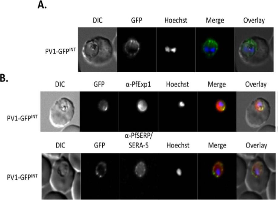
Live cell imaging. (A) Left: live cell imaging of the PV1-GFPINT transgenic line. (B) Upper panel, colocalisation of PV1-GFP chimera with PfExp1. Lower panel, colocalisation of PV1-GFP chimera with PfSERA-5. In merge and overlay: GFP, green; specific antibody, red; Hoechst, blue. Immunofluorescence co-localisation using antibodies against the PV and PVM markers SERA-5 and Exp-1 respectively verified that this fluorescent signal represented the PV.Chu T, Lingelbach K, Przyborski JM. Genetic evidence strongly support an essential role for PfPV1 in intra-erythrocytic growth of P. falciparum. PLoS One. 2011 6(3):e18396.
See original on MMPMore information
| PlasmoDB | PF3D7_1129100 |
| GeneDB | PF3D7_1129100 |
| Malaria Metabolic Pathways | Localisation images Pathways mapped to |
| Previous ID(s) | PF11_0302 |
| Orthologs | PBANKA_0919100 , PCHAS_0927000 , PKNH_0927100 , PVP01_0929800 , PVX_092070 , PY17X_0920800 |
| Google Scholar | Search for all mentions of this gene |