PF3D7_1121000 palmitoyltransferase DHHC3 (DHHC3)
Disruptability [+]
| Species | Disruptability | Reference | Submitter | |
|---|---|---|---|---|
| P. falciparum 3D7 |
Refractory |
27060339 | Theo Sanderson, Wellcome Trust Sanger Institute | |
| P. falciparum 3D7 |
Possible |
USF piggyBac screen (Insert. mut.) | USF PiggyBac Screen | |
| P. berghei ANKA |
Possible |
RMgm-888 | Imported from RMgmDB | |
| P. berghei ANKA |
Refractory |
PlasmoGEM (Barseq) | PlasmoGEM | |
Mutant phenotypes [+]
| Species | Stage | Phenotype | Reference | Submitter |
|---|---|---|---|---|
| P. berghei ANKA | Asexual |
No difference |
RMgm-888 | Imported from RMgmDB |
| P. berghei ANKA | Ookinete |
Difference from wild-type |
RMgm-888
Normal ookinete production. (Light microscope)morphology of mature ookinetes is normal. (Slightly) reduced motility of mutant ookinetes |
Imported from RMgmDB |
| P. berghei ANKA | Oocyst |
Difference from wild-type |
RMgm-888
Reduced (10 fold) oocyst production |
Imported from RMgmDB |
| P. berghei ANKA | Sporozoite |
Difference from wild-type |
RMgm-888
Reduced number (4-fold) of hemolymph sporozoitesReduced number (25-fold) of salivary gland sporozoites.Gliding motility of salivary gland mutant sporozoites is severely impaired, with only 10% of sporozoites producing trails |
Imported from RMgmDB |
| P. berghei ANKA | Liver |
Difference from wild-type |
RMgm-888
Mice injected with 5,000 or 10,000 hemolymph sporozoites of the mutant failed to develop blood-stage parasitemia.Mice inoculated with 5,000 DHHC3-ko sporozoites failed to develop blood-stage parasitemia and upon injection with 10,000 sporozoites, only one out of three mice developed a blood-stage infection, with a delay of three days compared to wild-type |
Imported from RMgmDB |
Imaging data (from Malaria Metabolic Pathways)
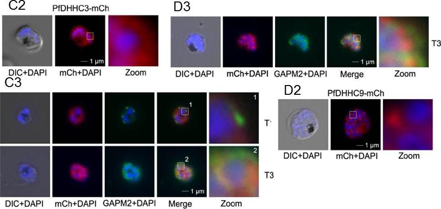
Over-expression and localization of PfDHHC3 and PFDHHC9 (PF11_0167) in late stage parasites. C. Expression of PfDHHC3-mCherry. C. Expression of PfDHHC3-mCherry. C2. Microscopic analysis located this GFP-fusion protein in the periphery of the nascent merozoites. C3. Co-localization with the IMC marker GAPM2 (anti-GAPM2, green) in fixed cells shows differential localization of PfDHHC2-mCherry with the IMC localization in early stages (T1) and colocalization in nascent merozoites (T3) consistent with plasma membrane association. Scale bar, 1 μm. D.Expression of PfDHHC9-mCherry. D2. Microscopy localized this fusion protein mainly in apical structures in the parasite. D3. Co-localization with the IMC marker GAPM2 (anti-GAPM2, green) in fixed cells shows differential localization of PfDHHC9 with the IMC (T3). Scale bar, 1 μm.Wetzel J, Herrmann S, Swapna LS, Prusty D, Peter AT, Kono M, Saini S, Nellimarla S, Wong TW, Wilcke L, Ramsay O, Cabrera A, Biller L, Heincke D, Mossman K, Spielmann T, Ungermann C, Parkinson J, Gilberger TW. The role of palmitoylation for protein recruitment to the inner membrane complex of the malaria parasite. J Biol Chem. 2014 Nov 25.
See original on MMP
Expression and localisation of PfDHHC proteins in P. falciparum schizonts. Triple-HA-tagged PfDHHC proteins were localised by immunofluorescence using antibodies against the 3-HA tag (green). Immunofluorescence staining of each of the tagged PfDHHC proteins was compared against that of the following known localisation markers (red): ERD2 (Golgi marker), BIP (endoplasmic reticulum marker), RAP1 (rhoptry marker), GAP45 (inner membrane complex marker) and MSP1 (plasma membrane marker). Nuclear staining by DAPI is shown in blue. For the staining of PfDHHC9 with GAP45 and MSP1, both a late schizont, as well as an early schizont, is shown in order to differentiate between IMC and plasma membrane localisation. Scale bar: 5 μm.Tay CL, Jones ML, Hodson N, Theron M, Choudhary JS, Rayner JC. Study of Plasmodium falciparum DHHC palmitoyl-transferases identifies a role for PfDHHC9 in gametocytogenesis. Cell Microbiol. 2016 Apr 6.
See original on MMP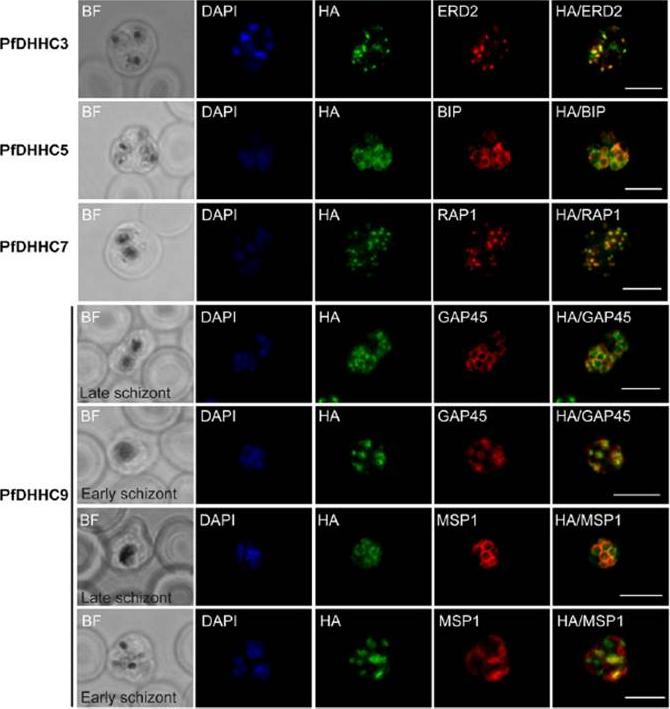
Expression and localization of PfDHHC proteins in Plasmodium falciparum schizonts. Triple-HA-tagged PfDHHC proteins were localized by immuno-fluorescence using antibodies against the 3-HA tag (green). Immunofluorescence staining of each of the tagged PfDHHC proteins was compared against that of the following known localization markers (red): ERD2 (Golgi marker), BIP (endoplasmic reticulum marker), RAP1 (rhoptry marker), GAP45 (inner membrane complex marker) and MSP1 (plasma membrane marker). Nuclear staining by DAPI is shown in blue. For the staining of PfDHHC9 with GAP45 and MSP1, a late schizont, as well as an early schizont, is shown in order to differentiate between inner membrane complex and plasma membrane localization. Scale bar: 5 μm.Tay CL, Jones ML, Hodson N, Theron M, Choudhary JS, Rayner JC. Study of Plasmodium falciparum DHHC palmitoyl transferases identifies a role for PfDHHC9 in gametocytogenesis. Cell Microbiol. 2016 18(11):1596-1610.
See original on MMPMore information
| PlasmoDB | PF3D7_1121000 |
| GeneDB | PF3D7_1121000 |
| Malaria Metabolic Pathways | Localisation images Pathways mapped to |
| Previous ID(s) | PF11_0217 |
| Orthologs | PBANKA_0927300 , PCHAS_0917000 , PKNH_0918700 , PVP01_0921600 , PVX_091670 , PY17X_0929300 |
| Google Scholar | Search for all mentions of this gene |