PF3D7_1108400 casein kinase 2, alpha subunit (CK2alpha)
Disruptability [+]
| Species | Disruptability | Reference | Submitter | |
|---|---|---|---|---|
| P. falciparum 3D7 |
Refractory |
22127061 | Theo Sanderson, Wellcome Trust Sanger Institute | |
| P. falciparum 3D7 |
Refractory |
USF piggyBac screen (Insert. mut.) | USF PiggyBac Screen | |
| P. falciparum 3D7 |
Refractory |
19114502 | Theo Sanderson, Francis Crick Institute | |
| P. berghei ANKA |
Refractory |
RMgm-558 | Imported from RMgmDB | |
| P. berghei ANKA |
Refractory |
PlasmoGEM (Barseq) | PlasmoGEM | |
Mutant phenotypes [+]
| Species | Stage | Phenotype | Reference | Submitter |
|---|---|---|---|---|
| P. falciparum 3D7 | Asexual |
Invasion defect |
26694741 (Knock down)
\" Depletion of PfCK2, a kinase implicated to phosphorylate these cytoplasmic tails, blocks P. falciparum invasion of red blood cells.\" |
Theo Sanderson, Francis Crick Institute |
| P. falciparum 3D7 | Gametocyte |
Difference from wild-type |
33712726 (Conditional)
\"Conditional knockdown of PfCK2α expression prevented the transition of stage IV into transmission-competent stage V gametocytes, whereas the conditional knockout of pfck2a completely blocked gametocyte maturation already at an earlier stage of sexual differentiation.\" |
Theo Sanderson, Francis Crick Institute |
Imaging data (from Malaria Metabolic Pathways)
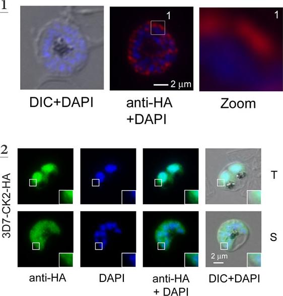
1. In vivo phosphorylation of S3233 (S3233 is the sole phosphoacceptor site in the cytoplasmic domain of Rh2B for in vitro phosphorylation by parasite extract) in schizonts . Expression was verified by IFA using anti-HA antibodies. Scale bar indicate 2 μm. Rh2B is localized at the apical pole of nascent merozoites.2. Expression, purification of PfCK2 alpha (endogenously HA-tagged (3D7-CK2-HA, PF3D7_1108400) and Rh2b in vitro phosphorylation. Immuno-fluorescence microscopy using endogenous derived PfCK2-HA alpha. Localization with the anti-HA antibodies (green) confirmed expression in the nucleus (blue, DAPI) and cytosol of fixed parasites in the trophozoite (T) and schizont (S) stage. Scale bar indicates 2 μm. Enlargements of selected areas are marked with a white square the endogenous tagged protein was localized in the parasite´s cytosol, as well as in the nucleus of trophozoite stage parasites. In the schizont stage of the parasite the dual localization of endogenous tagged CK2 was less pronounced and appeared predominantly cytosolic.Engelberg K, Paul AS, Prinz B, Kono M, Ching W, Heincke D, Dobner T, Spielmann T, Duraisingh M, Gilberger TW. Specific phosphorylation of the PfRh2b invasion ligand of Plasmodium falciparum. Biochem J. 2013 452(3):457-66
See original on MMP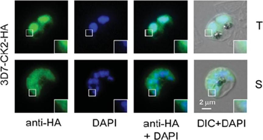
Immunofluorescence microscopy using endogenously derived Pf CK2α–HA. Localization with the anti-HA antibodies (green) confirmed expression in the nucleus (blue, DAPI) and cytosol of fixed parasites in the trophozoite (T) and schizont (S) stage. Scale bar indicates 2 μm. Enlargements of selected areas are marked with a white square. DIC, differential interference contrast microscopy. the endogenously tagged protein was localized in the parasite’s cytosol, as well as in the nucleus of trophozoite stage parasites. In the schizont stage of the parasite the dual localization of endogenously tagged CK2 was less pronounced and appeared predominantly cytosolic.Engelberg K, Paul AS, Prinz B, Kono M, Ching W, Heincke D, Dobner T, Spielmann T, Duraisingh MT, Gilberger TW. Specific phosphoryla tion of the PfRh2b invasion ligand of Plasmodium falciparum. Biochem J. 2013 452(3):457-66. PMID:
See original on MMP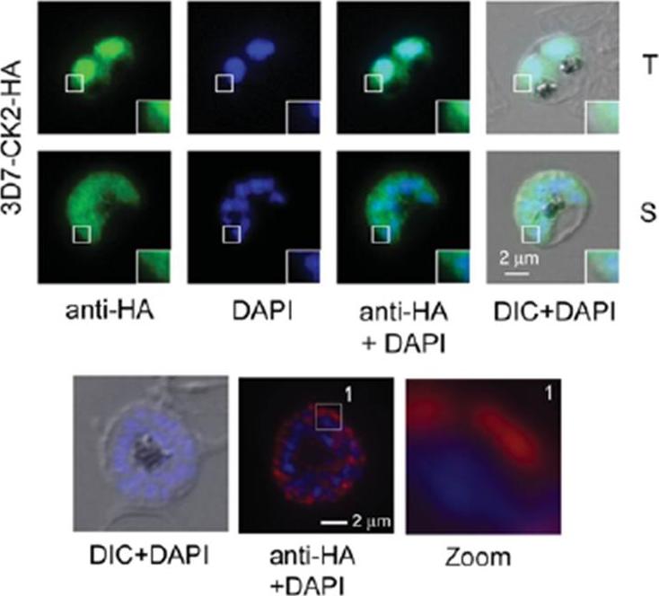
Upper pannel: Immunofluorescence microscopy using endogenously derived Pf CK2α–HA. Localization with the anti-HA antibodies (green) confirmedexpression in the nucleus (blue, DAPI) and cytosol of fixed parasites in the trophozoite (T) and schizont (S) stage. Scale bar indicates 2 μm. Enlargements of selected areas are marked with a white square. DIC, differential interference contrast microscopy. PF3D7_1108400. The endogenously tagged protein was localized in the parasite’s cytosol, as well as in the nucleus of trophozoite stage parasites. In the schizont stage of the parasite the dual localization ofendogenously tagged CK2 was less pronounced and appeared predominantly cytosolic. Lowe pannel: Expression of Rh2b–HA was verified by IFA using anti-HA antibodies. Scale bar, 2 μm. DIC, differential interference contrast microscopy. Rh2b–HA is localized at the apical pole of nascent merozoites.Engelberg K, Paul AS, Prinz B, Kono M, Ching W, Heincke D, Dobner T, Spielmann T, Duraisingh MT, Gilberger TW. Specific phosphorylation of the PfRh2b invasion ligand of Plasmodium falciparum. Biochem J. 2013 Jun 15;452(3):457-66.
See original on MMPMore information
| PlasmoDB | PF3D7_1108400 |
| GeneDB | PF3D7_1108400 |
| Malaria Metabolic Pathways | Localisation images Pathways mapped to |
| Previous ID(s) | PF11_0096 |
| Orthologs | PBANKA_0938600 , PCHAS_0905700 , PKNH_0906000 , PVP01_0909200 , PVX_091095 , PY17X_0941100 |
| Google Scholar | Search for all mentions of this gene |