PF3D7_1108200 D-tyrosyl-tRNA(Tyr) deacylase (DTD)
Disruptability [+]
| Species | Disruptability | Reference | Submitter |
|---|---|---|---|
| P. falciparum 3D7 |
Refractory |
USF piggyBac screen (Insert. mut.) | USF PiggyBac Screen |
Mutant phenotypes [+]
None reported yet. Please press the '+' button above to add one.Imaging data (from Malaria Metabolic Pathways)
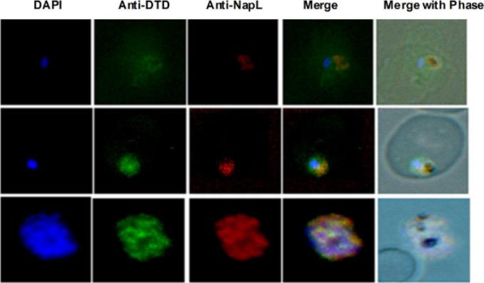
Expression and activity of PfDTD. Localization of PfDTD in different intra-erythrocytic stages of P. falciparum by immunofluo-rescence staining, Ring stage (R), trophozoite stage (T), and schizont stage (S) are shown. In each panel are shown: (i) image of cell stained with DAPI (blue), (ii) anti-PfDTD antibodies, (iii) anti-PfNapL antibodies, (iv) merged image, (v) merge with phase contrast. Results clearly show the colocalization of PfDTD with PfNapL at all the asexual life stages of parasite, suggesting PfDTD to be cytoplasmic in nature, like for PfNapL.Bhatt TK, Yogavel M, Wydau S, Berwal R, Sharma A. Ligand-bound structures provide atomic snapshots for the catalytic mechanism of D-amino acid deacylase. J Biol Chem. 2010 Feb 285(8):5917-30.
See original on MMP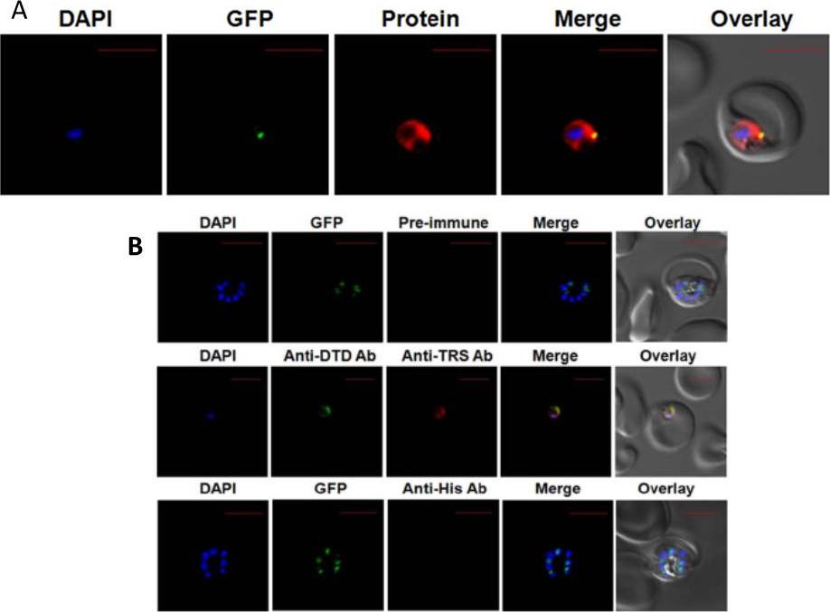
Expression and localization of tRNAThr synthetase (Pf-Ed-TRS). A. Displays cellular localizations. Data clearly show in the parasite apicoplast as well as in the cytoplasm. B. Upper and lower panels show confocal IFA with pre-immune sera and with anti-histidine antibodies, whereas the middle panels depict cytoplasmic staining of Pf-DTD. The parasite line used was GFP-tagged (strain D10 ACPleader-GFP) where apicoplast fluoresces green. DAPI staining is in blue while aminoacyl-tRNA synthetases are stained with Alexa594 (red).Khan S, Sharma A, Jamwal A, Sharma V, Pole AK, Thakur KK, Sharma A. Uneven spread of cis- and trans-editing aminoacyl-tRNA synthetase domains within translational compartments of P. falciparum. Sci Rep. 2011;1:188.
See original on MMP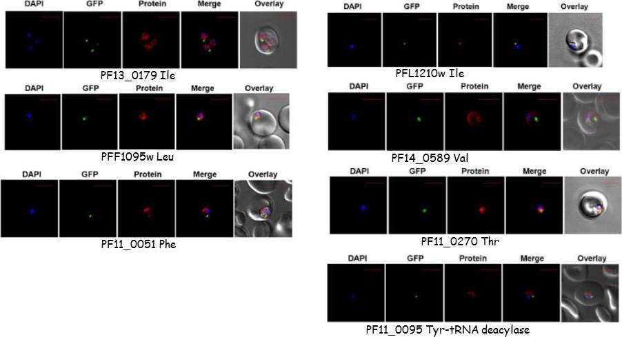
Targeting of P. falciparum aminoacyl-tRNA synthetase and of DTD. The parasite line used was GFP-tagged (strain D10 ACPleader-GFP) where apicoplast fluorescence is in green. DAPI staining is in blue while aminoacyl-tRNA synthetases are stained with Alexa594 (red). only the late ring and early trophozoite-stage localizations are depicted here but patterns were identical in other intra-erythrocytic stages. All tRNA synthases were cytoplasmic except PFL1210w Ile which is seen located in the apicoplast and deacylase which is mitochondrial.Khan S, Sharma A, Jamwal A, Sharma V, Pole AK, Thakur KK, Sharma A. Uneven spread of cis- and trans-editing aminoacyl-tRNA synthetase domains within translational compartments of P. falciparum. Sci Rep. 2011;1:188.
See original on MMP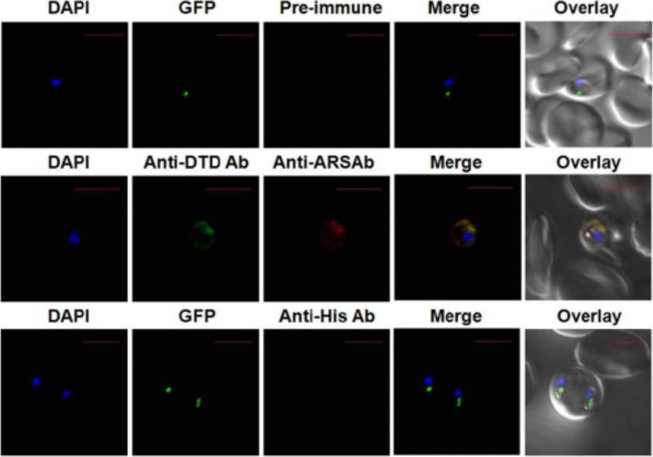
Expression and localization of tRNAAla synthetase (Pf-Ed-ARS). Displays cellular localizations. Upper and lower panels show confocal IFA with pre-immune sera and with anti-histidine antibodies, whereas the middle panel depicts cytoplasmic staining of Pf-DTD. Data clearly show in the parasite apicoplast as well as in the cytoplasm. The parasite line used was GFP-tagged (strain D10 ACPleader-GFP) where apicoplast fluoresces green. DAPI staining is in blue while aminoacyl-tRNA synthetases are stained with Alexa594 (red).Khan S, Sharma A, Jamwal A, Sharma V, Pole AK, Thakur KK, Sharma A. Uneven spread of cis- and trans-editing aminoacyl-tRNA synthetase domains within translational compartments of P. falciparum. Sci Rep. 2011;1:188.
See original on MMP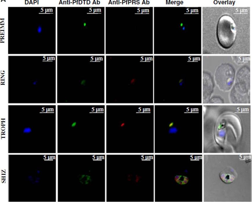
Sub-cellular localization of PfPRS. Confocal microscopy of Plasmodium falciparum localization in asexual parasite stages—rings, trophozoites and schizonts using anti-sera raised against PfPRS254-746. Pf D-tyrosyltRNA Tyr deacylase (PfDTD) was used as a marker for parasite cytosol. Parasite nucleus was stained with DAPI (blue) while PfPRS and PfDTD were stained with Alexa Fluor 594 (red) and Alexa Fluor 488 (green) respectively. Scale bar in white corresponds to 5 mm size. Pre-immune serum was used as negative control in these experiments. PfPRS seemed to be present only in the parasite cytoplasm within ring, trophozoite and schizont stages .This suggests that PfPRS contributes to production of prolyladenylate in parasite cytoplasm, and is distinct from the apicoplastic (PfPRSapi). PfPRS seems confined to parasite cytoplasm in asexual blood stages of the parasite, and is not secreted like Plasmodium falciparum tyrosyl-tRNA synthetase (PfYRS).Jain V, Kikuchi H, Oshima Y, Sharma A, Yogavel M. Structural and functional analysis of the anti-malarial drug target prolyl-tRNA synthetase. J Struct Funct Genomics. 2014 15(4):181-90.
See original on MMP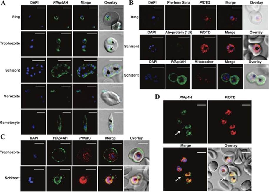
Spatial distribution of P. falciparum Ap4AH during erythrocytic schizogony. Shown are DAPI staining of nucleus in blue and PfAp4AH stained with Alexa 488 in green. (A) Confocal microscopy-data based spatial distribution of PfAp4AH in infected RBCs. PfAp4AH is non-nuclear in blood stages of the parasite and resides in its cytoplasm. (B) Non-mitochondrial localization with various controls is shown. Upper panel shows pre-immune serum (Pre-Imm Sera) control which does not stain the parasite or RBCs. Middle panel shows competitive binding of anti-PfAp4AH antibody to infected cells, where anti-PfAp4AH antibodies were incubated with recombinant PfAp4AH protein in 1:5 ratio. Pf D-tyrosyl-tRNATyr deacylase (DTD) is a cytoplasmic marker. Lower panel shows non-mitochondrial localization where mtochondria are stained in red. (C) RBC membrane localization of PfAp4AH during trophozoite and schizont stages of parasite. VarC is a marker for infected RBC membrane localization. (D) A field view of anti-PfAp4AH antibody staining of infected RBCs. Significant fraction of cells (~50%) showed membrane localization of PfAp4AH - here cell is marked with white arrow. Uninfected RBCs (without DAPI and PfDTD staining here) are unstained. White scale bar in confocal figures is of 5 μmSharma A, Yogavel M, Sharma A. Structural and functional attributes of malaria parasite diadenosine tetraphosphate hydrolase. Sci Rep. 2016 Feb 1;6:19981.
See original on MMPMore information
| PlasmoDB | PF3D7_1108200 |
| GeneDB | PF3D7_1108200 |
| Malaria Metabolic Pathways | Localisation images Pathways mapped to |
| Previous ID(s) | PF11_0095 |
| Orthologs | PBANKA_0938700 , PCHAS_0905600 , PKNH_0905800 , PVP01_0909000 , PVX_091090 , PY17X_0941200 |
| Google Scholar | Search for all mentions of this gene |