PF3D7_1036000 merozoite surface protein (MSP11)
Mutant phenotypes [+]
| Species | Stage | Phenotype | Reference | Submitter |
|---|---|---|---|---|
| P. falciparum 3D7 | Asexual |
No difference |
15664649 | Theo Sanderson, Wellcome Trust Sanger Institute |
Imaging data (from Malaria Metabolic Pathways)
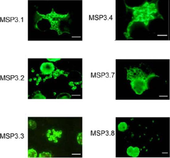
Expression analysis of MSP3-like ORFs. IFA analysis of acetone-fixed thin smear of the blood stage parasites, using the same antibodies used for Western blot analysis, shows merozoite surface staining. The size-bar drawn in the lower right-hand corner of each microscopic field represents 5 mm. Antibodies affinity-purified against the unique region of MSP3.5 did not react to parasite proteins in IFA.Singh S, Soe S, Weisman S, Barnwell JW, Pérignon JL, Druilhe P. A conserved multi-gene family induces cross-reactive antibodies effective in defense against Plasmodium falciparum. PLoS One. 2009;4(4):e5410.
See original on MMP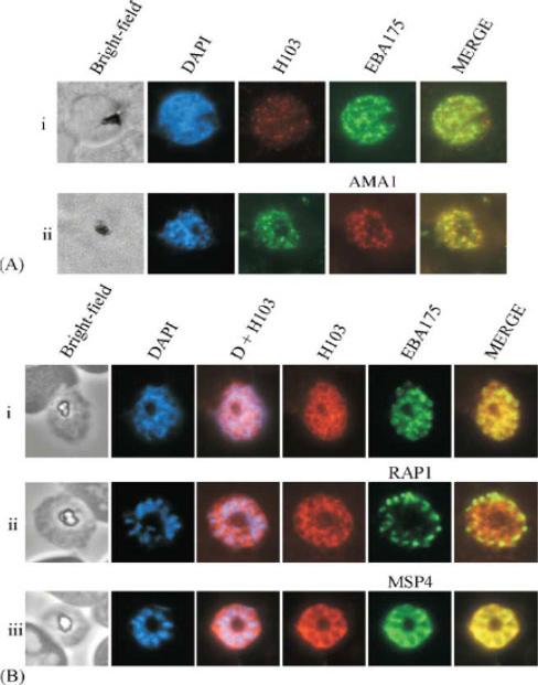
(A) Immunofluorescence of early schizont parasites indicated that H103 partially co-localises with EBA175 (i) and AMA1 (ii) in the micronemes. DAPI staining indicates that the parasites are early schizonts. (B) Immunofluorescence of late stage schizont parasites using antibodies to H103, EBA175 (i), RAP1 (ii) and MSP4 (iii) indicated that at this stage of the asexual cycle H103 was located on the merozoite surface. H103 did not co-localise with the microneme protein EBA175 or the rhoptries protein RAP1 at this stage of the cycle. DAPI staining shows the location of DNA and signifies that the parasites are fully segmented schizonts.Pearce JA, Mills K, Triglia T, Cowman AF, Anders RF. Characterisation of two novel proteins from the asexual stage of Plasmodium falciparum, H101 and H103. Mol Biochem Parasitol. 2005 139:141-51. Copyright Elsevier 2009.
See original on MMP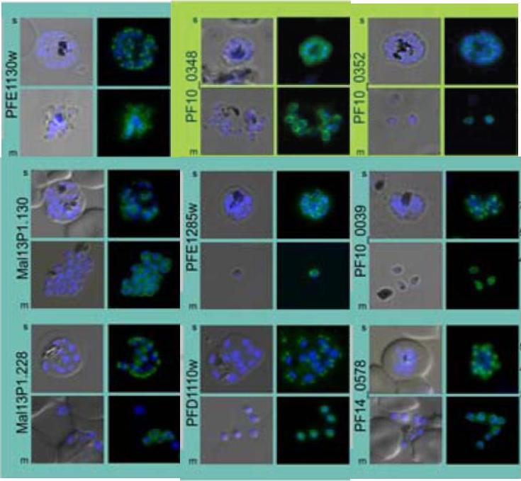
Subcellular distribution of proteins predicted to be involved in invasion. All proteins were localized in schizonts (s) and free merozoites (m) using GFP-fusion proteins and grouped according to their predominant GFP localization: surface (green), inner membrane complex (turquoise),Hu G, Cabrera A, Kono M, Mok S, Chaal BK, Haase S, Engelberg K, Cheemadan S, Spielmann T, Preiser PR, Gilberger TW, Bozdech Z. Transcriptional profiling of growth perturbations of the human malaria parasite Plasmodium falciparum. Nat Biotechnol. 2010 28(1):91-8.
See original on MMPMore information
| PlasmoDB | PF3D7_1036000 |
| GeneDB | PF3D7_1036000 |
| Malaria Metabolic Pathways | Localisation images Pathways mapped to |
| Previous ID(s) | PF10_0352 |
| Orthologs | |
| Google Scholar | Search for all mentions of this gene |