PF3D7_1035700 duffy binding-like merozoite surface protein (DBLMSP)
Disruptability [+]
| Species | Disruptability | Reference | Submitter |
|---|---|---|---|
| P. falciparum 3D7 |
Possible |
22986493 | Theo Sanderson, Wellcome Trust Sanger Institute |
| P. falciparum 3D7 |
Possible |
USF piggyBac screen (Insert. mut.) | USF PiggyBac Screen |
Mutant phenotypes [+]
None reported yet. Please press the '+' button above to add one.Imaging data (from Malaria Metabolic Pathways)
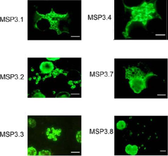
Expression analysis of MSP3-like ORFs. IFA analysis of acetone-fixed thin smear of the blood stage parasites, using the same antibodies used for Western blot analysis, shows merozoite surface staining. The size-bar drawn in the lower right-hand corner of each microscopic field represents 5 mm. Antibodies affinity-purified against the unique region of MSP3.5 did not react to parasite proteins in IFA.Singh S, Soe S, Weisman S, Barnwell JW, Pérignon JL, Druilhe P. A conserved multi-gene family induces cross-reactive antibodies effective in defense against Plasmodium falciparum. PLoS One. 2009;4(4):e5410.
See original on MMP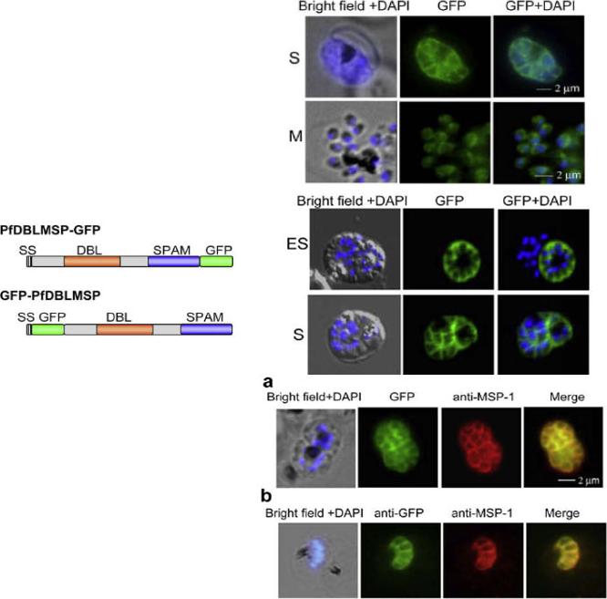
(D, E) Sub-cellular distribution of of PfDBLMSP on the merozoite surface. Fluorescent microscopic images of live parasites expressing PfDBLMSPGFP (D) and GFP-PfDBLMSP (E) fusion proteins. Both the fusion proteins (green) are localized on the surface of the merozoites. The nucleus is stained with DAPI (blue). ES, early schizont; S, schizont; and M, Merozoites. (F) Co-localization studies of GFP fusion proteins with the surface marker MSP-1 PFI1475w by immunofluorescence. The PfDBLMSP-GFP (a) and GFP-PfDBLMSP (b) fusion proteins (green) co-localize with the surface protein MSP-1 (red) as shown in the merge picture. Nuclei are stained with DAPI (blue).Schematic domain structure of PfDBLMSP as N- or C-terminal GFP fusion, labeled as PfDBLMSP-GFP and GFP-PfDBLMSP, respectively. Signal peptides: black; green fluorescent protein: green; Duffy binding like (DBL) domain: brown; secreted polymorphic antigen associated with merozoites (SPAM) domain: blue.Wickramarachchi T, Cabrera AL, Sinha D, Dhawan S, Chandran T, Devi YS, Kono M, Spielmann T, Gilberger TW, Chauhan VS, Mohmmed A. A novel Plasmodium falciparum erythrocyte binding protein associated with the merozoite surface, PfDBLMSP. Int J Parasitol. 2009 39:763-73. Copyright Elsevier 2009.
See original on MMP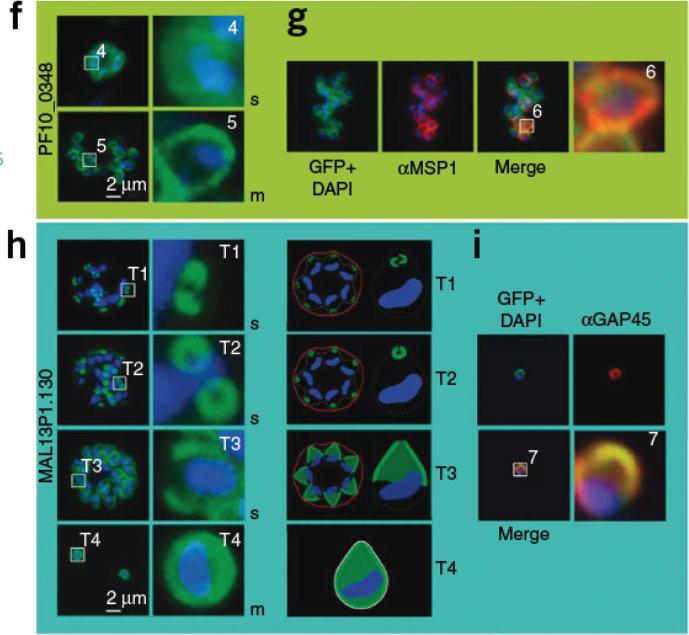
Boxed regions are numbered and depicted in higher magnification to the right. The nucleus is stained with DAPI (blue). f) PF10_0348-GFP (green) localized to the surface of schizonts and free merozoites in unfixed parasites. g) PF10_0348-GFP co-localized with the surface protein MSP-1 (red) in fixed parasites. Dynamics of MAL13P1.130-GFP (green) during schizogony in unfixed parasites. In early schizogony (T1), MAL13P1.130-GFP emerged as a cramp-like-structure at the apical tip of forming merozoites (h). This structure develops to be ring-like (T2) before becoming evenly distributed at the periphery of the nascent merozoite (T3-4). The third row shows a schematic representation. For confocal three-dimensional reconstitution,Hu G, Cabrera A, Kono M, Mok S, Chaal BK, Haase S, Engelberg K, Cheemadan S, Spielmann T, Preiser PR, Gilberger TW, Bozdech Z. Transcriptional profiling of growth perturbations of the human malaria parasite Plasmodium falciparum. Nature Biotechnol. 2010 28:91-8.
See original on MMP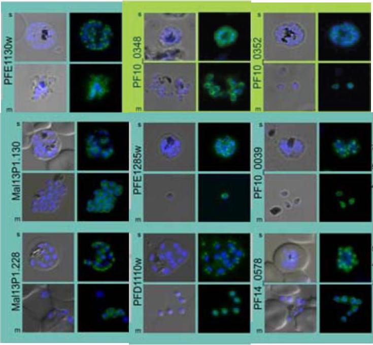
Subcellular distribution of proteins predicted to be involved in invasion. All proteins were localized in schizonts (s) and free merozoites (m) using GFP-fusion proteins and grouped according to their predominant GFP localization: surface (green), inner membrane complex (turquoise),Hu G, Cabrera A, Kono M, Mok S, Chaal BK, Haase S, Engelberg K, Cheemadan S, Spielmann T, Preiser PR, Gilberger TW, Bozdech Z. Transcriptional profiling of growth perturbations of the human malaria parasite Plasmodium falciparum. Nat Biotechnol. 2010 28(1):91-8.
See original on MMP
Processed Peripheral MSPs become part of an anchored complex late in schizogony. Immunofluorescence assays of Trophozoites (28-32 hr) in columns 1 (Phase + 488 + DAPI) and 2 (488 + DAPI), Schizonts (44-48 hr) in columns 3 (Phase + 488 + DAPI) and 4 (488 + DAPI) and PEMS (E64 treated) in columns 5 (Phase + 488 + DAPI) and 6 (488 + DAPI) were probed with monoclonal antibodies (green); 9A6 (anti-MSP1 p83), 9A12 (anti-MSP3), 2C12 (anti-MSP6), 7H12 (anti-MSPDBL1), 4A7 (anti-MSPDBL2) and 8F1 (anti-MSP7). EXP2 was used as an expression control probed with polyclonal rabbit anti-EXP2 antibodies (red). The nuclei of parasites are stained with DAPI (blue).Lin CS, Uboldi AD, Epp C, Bujard H, Tsuboi T, Czabotar PE, Cowman AF. Multiple P. falciparum Merozoite Surface Protein 1 Complexes Mediate Merozoite Binding to Human Erythrocytes. J Biol Chem. 2016 Jan 28. [Epub ahead of print] PMID:
See original on MMP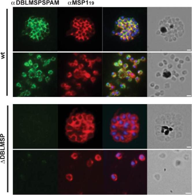
Indirect immunofluorescence assay on thin smears of schizont and merozoites from wild-type and Δdblmsp parasites showing expression of DBLMSP (green; first panel) and MSP1 (red; second panel). Third panel depicts the overlay of both antibodies with the nuclear stain DAPI; forth panel shows the corresponding brightfield images. Size bars represent 1μm.Crosnier C, Iqbal Z, Knuepfer E, Maciuca S, Perrin AJ, Kamuyu G, Goulding D, Bustamante LY, Miles A, Moore SC, Dougan G, Holder AA, Kwiatkowski DP, Rayner JC, Pleass RJ, Wright GJ. Binding of Plasmodium falciparum merozoite surface proteins DBLMSP and DBLMSP2 to human immunoglobulin M is conserved amongst broadly diverged sequence variants. J Biol Chem. 2016 May 12. [Epub ahead of print] PMID:
See original on MMPMore information
| PlasmoDB | PF3D7_1035700 |
| GeneDB | PF3D7_1035700 |
| Malaria Metabolic Pathways | Localisation images Pathways mapped to |
| Previous ID(s) | PF10_0348 |
| Orthologs | |
| Google Scholar | Search for all mentions of this gene |