PF3D7_1035400 merozoite surface protein 3 (MSP3)
Disruptability [+]
Mutant phenotypes [+]
| Species | Stage | Phenotype | Reference | Submitter |
|---|---|---|---|---|
| P. falciparum 3D7 | Asexual |
Attenuated |
11952894 (Knock down)
truncation -> mistargetomg |
Theo Sanderson, Wellcome Trust Sanger Institute |
| P. falciparum 3D7 | Asexual |
Enhanced virulence |
28484054 Enhanced invasion in the presence of complement |
Theo Sanderson, Wellcome Trust Sanger Institute |
Imaging data (from Malaria Metabolic Pathways)
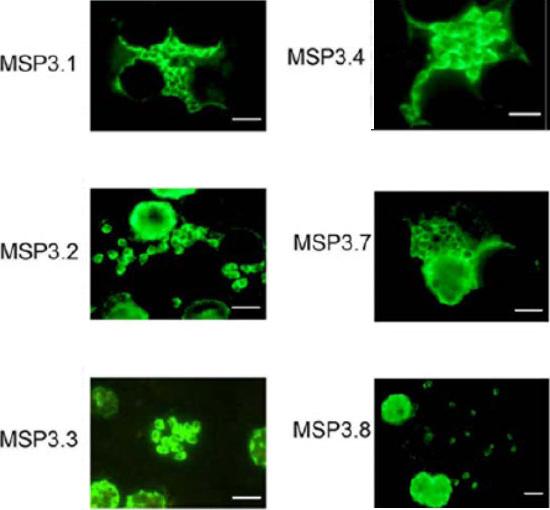
Expression analysis of MSP3-like ORFs. IFA analysis of acetone-fixed thin smear of the blood stage parasites, using the same antibodies used for Western blot analysis, shows merozoite surface staining. The size-bar drawn in the lower right-hand corner of each microscopic field represents 5 mm. Antibodies affinity-purified against the unique region of MSP3.5 did not react to parasite proteins in IFA.Singh S, Soe S, Weisman S, Barnwell JW, Pérignon JL, Druilhe P. A conserved multi-gene family induces cross-reactive antibodies effective in defense against Plasmodium falciparum. PLoS One. 2009;4(4):e5410.
See original on MMP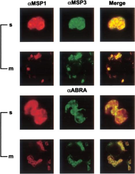
Subcellular localization of MSP3 and ABRA Smears of late schizonts (s) of anti-MSP1 (located on the surface of the merozoite (m)) and anti-MSP3 or anti-ABRA antibodies. Anti-MSP1 mouse antibodies were labeled with rhodamine-conjugated anti-mouse antibodies (red). Anti-MSP3 and anti-ABRA rabbit antibodies were labeled with anti-rabbit antibodies conjugated with FITC (green). Co-localization is shown in the row and indicated by yellow. D10 smears labeled with anti-MSP1 (first column) and anti-MSP-3 or anti-ABRA (second column). In late D10 schizonts, MSP-3, MSP-1 and ABRA had a localization consistent with the parasitophorous vacuole and the merozoite surface. Mills KE, Pearce JA, Crabb BS, Cowman AF. Truncation of merozoite surface protein 3 disrupts its trafficking and that of acidic-basic repeat protein to the surface of Plasmodium falciparum merozoites. Mol Microbiol. 2002 43:1401-1411. Copyright John Wiley & Sons Ltd. 2010.
See original on MMP
(B–D) Immunofluorescence microscopy of fixed smears of P. falciparum parasites using anti-PfStx1 antiserum. Secondary antisera (Molecular Probes) labeled with a fluorophore were used for visualization on an Olympus BX60 fluorescence microscope, and images were merged with Adobe Photoshop 5.0. In trophozoite stage parasites PfStx1 (red) has a plasma membrane like staining pattern and does not appear to be exported into the erythrocyte (B). In schizont stage parasites, PfStx1 (green) again has a peripheral location that is distinct from the ER marker PfBiP PFI0875w (in red) (C). PfStx1 (red) co-localizes with PfMSP3 (in green), a plasma membrane marker in schizont stage parasites (D), confirming its predicted location proximal to the plasma membrane.Parish LA, Rayner JC. Plasmodium falciparum secretory pathway: Characterization of PfStx1, a plasma membrane Qa-SNARE. Mol Biochem Parasitol. 2009 164:153-6. Copyright Elsevier 2009. PMID:
See original on MMP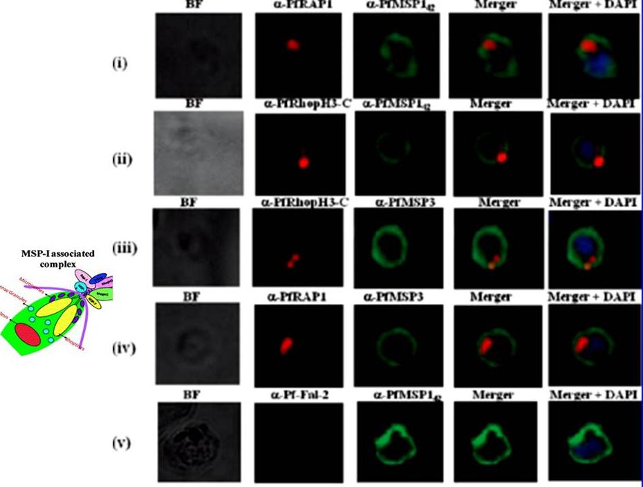
Localization of proteins of PfMSP-1 associated complex by immunofluorescence assay. Unpermeabilized P. falciparum merozoites were coimmunostained with anti-PfRAP-1 and anti-PfMSP-142 antibodies (i), with anti-PfRhopH3-C and anti-PfMSP-142 antibodies (ii), with anti-PfRhopH3-C and anti-PfMSP-3 antibodies (iii), anti-PfRAP-1 and anti-PfMSP-3 antibodies (iv), and anti-Pffalcipain-2 and anti-PfMSP-142 antibodies (v). PfMSP-1 and PfMSP-3 staining was detected on the entire surface of merozoites, while PfRhopH3 and PfRAP-1 staining localized at the apical end of merozoites. Co-localization was observed between PfMSP-1 and PfRAP-1, PfMSP-1 and PfRhopH3, PfMSP-3 and PfRhopH3 as well PfMSP-1 and PfMSP-3 staining was detected on the entire surface of merozoites, while PfRhopH3 and PfRAP-1 staining localized at the apical end of merozoites. Co-localization was observed between PfMSP-1 and PfRAP-1, PfMSP-1 and PfRhopH3, PfMSP-3 and PfRhopH3 as well antibody and the specific location of PfRhopH3 in rhoptries. Ranjan R, Chugh M, Kumar S, Singh S, Kanodia S, Hossain MJ, Korde R, Grover A, Dhawan S, Chauhan VS, Reddy VS, Mohmmed A, Malhotra P. Proteome analysis Rrveals a large merozoite surface protein-1 associated complex on the Plasmodium falciparum merozoite surface. J Proteome Res. 2010 10:680-91. Copyright American Chemical Society
See original on MMP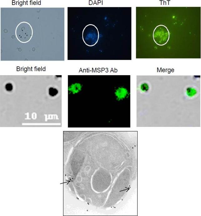
A. Thioflavin-T staining of egressed schizonts along with DAPI suggests presence of amyloid coat on the merozoite surface. (B) Confocal assay visualizing hemozoin crystals stained by anti-MSP3 Abs.(C) Immuno electron microscopy using anti–MSP3 Abs showed peripheral localization of MSP3 over merozoite surface. Merozoites immunostained with anti-MSP3 Abs revealed novel long fibrillar assemblies associated with the surface, possessing characteristics often attributed to amyloid like structures including fibril morphology and binding to ThT.Imam M, Singh S, Kaushik NK, Chauhan VS. Plasmodium falciparum Merozoite Surface Protein 3: oligomerization, self-assembly and heme complex formation. J Biol Chem. 2013 289(7):3856-68 PMID:
See original on MMP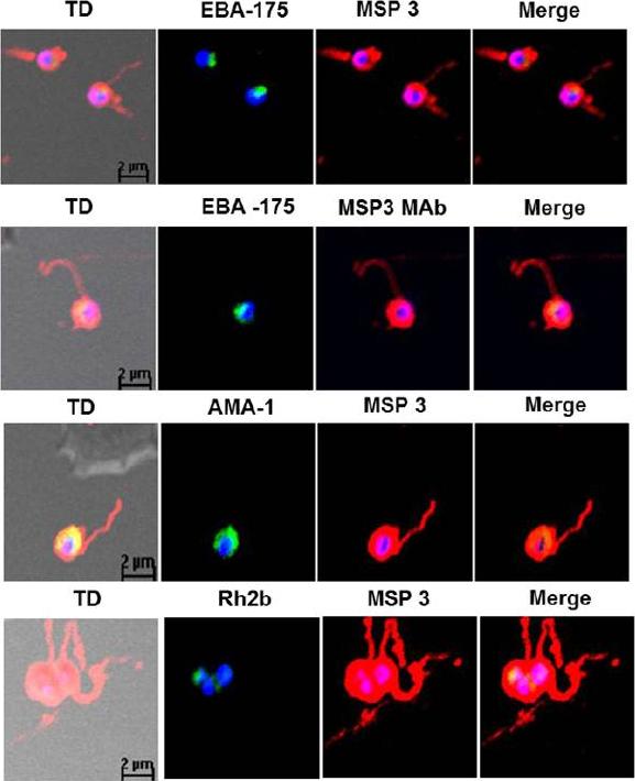
Detection of fibrillar structures in Plasmodium falciparum merozoites by immunofluorescence assay. Co-immunostaining of free merozoites with anti-MSP3 Abs along with Abs against EBA-175, AMA-1, Rh2b and MSP5. In all the slides long fibrous structure attached to free merozoite was detected by anti-MSP3 Abs. Monoclonal MSP3 antibodies also stained these fibrillar structures associated with merozoites, but other marker protein did not detect fibrillar assemblies. The long fibrils like structures attached at one end with merozoite surface were consistently seen to associate to distant erythrocytes as well as wrap around them.Imam M, Singh S, Kaushik NK, Chauhan VS. Plasmodium falciparum Merozoite Surface Protein 3: oligomerization, self-assembly and heme complex formation. J Biol Chem. 2013 289(7):3856-68
See original on MMP
Peripherally associated proteins MSP3, MSP7, SERA4, and SERA5 are shed during invasion. Invading merozoites were labeled with PfRON4 as a marker of the tight junction (green) and colabeled with antibodies directed against MSP3 (A), MSP7 (B), SERA4 (C), or SERA5 (D). All proteins were shed at the point of tight junction, with antibody reactivity to the merozoite surface being observed only on the proportion of the merozoite external to the RBC. All primary antibodies were used at a 1:100 serum dilution, and secondary antibodies were used at a 1:500 dilution.Boyle MJ, Langer C, Chan JA, Hodder AN, Coppel RL, Anders RF, Beeson JG. Sequential processing of merozoite surface proteins during and after erythrocyte invasion by Plasmodium falciparum. Infect Immun. 2014 82(3):924-36.
See original on MMP
Processed Peripheral MSPs become part of an anchored complex late in schizogony. Immunofluorescence assays of Trophozoites (28-32 hr) in columns 1 (Phase + 488 + DAPI) and 2 (488 + DAPI), Schizonts (44-48 hr) in columns 3 (Phase + 488 + DAPI) and 4 (488 + DAPI) and PEMS (E64 treated) in columns 5 (Phase + 488 + DAPI) and 6 (488 + DAPI) were probed with monoclonal antibodies (green); 9A6 (anti-MSP1 p83), 9A12 (anti-MSP3), 2C12 (anti-MSP6), 7H12 (anti-MSPDBL1), 4A7 (anti-MSPDBL2) and 8F1 (anti-MSP7). EXP2 was used as an expression control probed with polyclonal rabbit anti-EXP2 antibodies (red). The nuclei of parasites are stained with DAPI (blue).Lin CS, Uboldi AD, Epp C, Bujard H, Tsuboi T, Czabotar PE, Cowman AF. Multiple P. falciparum Merozoite Surface Protein 1 Complexes Mediate Merozoite Binding to Human Erythrocytes. J Biol Chem. 2016 Jan 28. [Epub ahead of print] PMID:
See original on MMPMore information
| PlasmoDB | PF3D7_1035400 |
| GeneDB | PF3D7_1035400 |
| Malaria Metabolic Pathways | Localisation images Pathways mapped to |
| Previous ID(s) | PF10_0345 |
| Orthologs | |
| Google Scholar | Search for all mentions of this gene |