PF3D7_1035200 S-antigen
Disruptability [+]
| Species | Disruptability | Reference | Submitter |
|---|---|---|---|
| P. falciparum 3D7 |
Refractory |
22986493 | Theo Sanderson, Wellcome Trust Sanger Institute |
| P. falciparum 3D7 |
Possible |
USF piggyBac screen (Insert. mut.) | USF PiggyBac Screen |
Mutant phenotypes [+]
None reported yet. Please press the '+' button above to add one.Imaging data (from Malaria Metabolic Pathways)
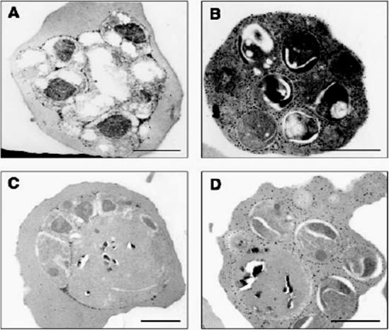
Detection of S-antigen by immunogold labeling with anti-S-antigen antibodies on ultra-thin sections of wild-type P. falciparum parasites. A, in untreated early schizonts, S-antigen localizes to the parasitophorous vacuole. B, in untreated late schizonts, S-antigen localizes throughout the erythrocyte cytosol. C, in leupeptin- and chymostatin-treated early schizonts, S-antigen localizes to the parasitophorous vacuole. D, in leupeptin- and chymostatin-treated late schizonts, S-antigen localizes throughout the erythrocyte cytosol. Scale bar 1.0 mm.Wickham ME, Culvenor JG, Cowman AF. Selective inhibition of a two-step egress of malaria parasites from the host erythrocyte. J Biol Chem. 2003 278:37658-63.
See original on MMP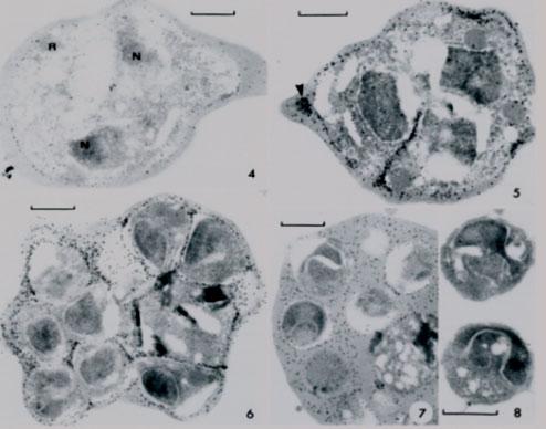
Electron micrographs from different parasite stages. 4. Early schizonts with 2 nuclei (N) and rhoprtry (R) and labeling over parasitophorous vacuole (PV). 5. Schizonts with labeled invaginating PV and with labeled vesicles within erythrocye cytoplasm (arrwohead). 6. Segmented schizont with labeled PV surrounding merozoites and residual body (containing pigment). Schizont with mature merozoites and PV membrane breakdown. Note even labeling of mixed RBC cytoplasm and PV contents. 8. Free merozoites in culture, free of specific label.Culvenor JG, Crewther PE. S-antigen localization in the erythrocytic stages of Plasmodium falciparum. J Protozool. 1990 37:59-65. Copyright John Wiley & Sons Ltd. 2010.
See original on MMP
Immunoelectron micrographs showing localisation of the S-antigen in 0.05% glutaraldehyde-fixed, parasitised erythrocytes, which were embedded in LR White resin (colloidal gold 10 nm). a Immunolabelling of the S-antigen is clearly visible within the parasitophorous vacuole, b No labelling is evident on the parasitised erythrocytic cytoplasm. M, merozoite; P, parasite; P VM, parasitophorous vacuole membrane. Bar = 0.5 mm.Ingram LT, Stenzel DJ, Kara UA, Bushell GR. Localisation of internal antigens of Plasmodium falciparum using monoclonal antibodies and colloidal gold. Parasitol Res. 1988;74:208-15.
See original on MMP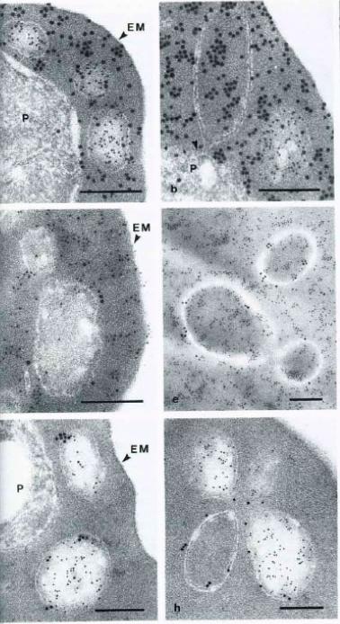
Immunoelectron micrographs of P. falciparum-infected erythrocytes showing double labeling results using anti-parasite and anti-host cell antibodies reacted with colloidal gold IgG of different particle sizes. A-B Abs directed against S-antigen (10 nm) and erythrocyte material (15 nm); D-E Abs against Exp-1 (15 nm) abd erythrocyte material (10 nm); G-H Abs against Exp-1 (15 nm) and against S-antigen (10 nm). EM – erythrocyte membrane; P – parasite; Arrowheads indicate pvm. Multiple species of vesicles, each with specifically packaged contents, are consistent with a sorting function of vesicular structures in the Plasmodium infected erythrocyte. During schizogony, two parasite antigens, an S-antigen and a parasitophorous vacuole membrane antigen, EXP-1, become packaged into such vesicles and are transported into the erythrocyte cytoplasm. At this stage of parasite development, host cell material is taken in through the parasitophorous vacuole membrane into the vacuolar space surrounding the parasite.Stenzel DJ, Kara UA. Sorting of malarial antigens into vesicular compartments within the host cell cytoplasm as demonstrated by immunoelectron microscopy. Eur J Cell Biol. 1989 49:311-8. Copyright Elsevier 2010.
See original on MMP
Detection of S-antigen by immunogold labeling with anti-S-antigen antibodies on ultra-thin sections of wild-type P. falciparum parasites. A, in untreated early schizonts, S-antigen localizes to the parasitophorous vacuole. B, in untreated late schizonts, S-antigen localizes throughout the erythrocyte cytosol. C, in leupeptin- and chymostatin-treated early schizonts, S-antigen localizes to the parasitophorous vacuole. D, in leupeptin- and chymostatin-treated late schizonts, S-antigen localizes throughout the erythrocyte cytosol. Scale bar 1.0 mm.Wickham ME, Culvenor JG, Cowman AF. Selective inhibition of a two-step egress of malaria parasites from the host erythrocyte. J Biol Chem. 2003 278:37658-63
See original on MMP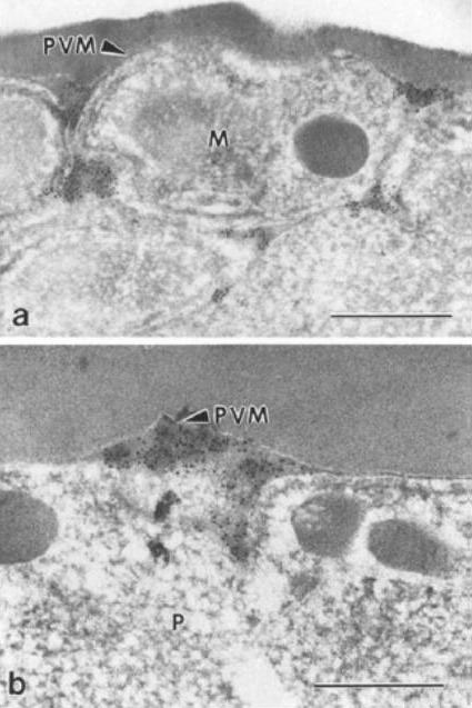
Immunoelectron micrographs showing localisation of the S-antigen in 0.05% glutaraldehyde-fixed, parasitised erythrocytes, which were embedded in LR White resin (colloidal gold 10 nm). a Immunolabelling of the S-antigen is clearly visible within the parasitophorous vacuole, b No labelling is evident on the parasitised erythrocytic cytoplasm. M, merozoite; P, parasite; P VM, parasitophorous vacuole membrane. Bar = 0.5 mm.Ingram LT, Stenzel DJ, Kara UA, Bushell GR. Localisation of internal antigens of Plasmodium falciparum using monoclonal antibodies and colloidal gold.Parasitol Res. 1988;74:208-15.
See original on MMPMore information
| PlasmoDB | PF3D7_1035200 |
| GeneDB | PF3D7_1035200 |
| Malaria Metabolic Pathways | Localisation images Pathways mapped to |
| Previous ID(s) | PF10_0343 |
| Orthologs | |
| Google Scholar | Search for all mentions of this gene |