PF3D7_1027700 centrin-3 (CEN3)
Disruptability [+]
| Species | Disruptability | Reference | Submitter |
|---|---|---|---|
| P. falciparum 3D7 |
Possible |
USF piggyBac screen (Insert. mut.) | USF PiggyBac Screen |
Mutant phenotypes [+]
None reported yet. Please press the '+' button above to add one.Imaging data (from Malaria Metabolic Pathways)

Right: Expression of P. falciparum centrin in schizonts. P. falciparum parasites were probed against mouse anti-PfCEN1, -2, -3, and -4 antibodies. Centrins are associated with the mitotic spindle.Left: Immunoelectron micrograph showing reactivity with the P. falciparum centrin-2 antibody. The gold particles associated with the nucleus of P. falciparum sporozoite stage parasite (A) and nucleus of asexual blood stage schizonts (B). N, nucleus; PVM, parasitophorous vacuole membrane; RBC, red blood cell. In sporozoites, IEM localization of PfCEN2 was observed as multiple dots in an area surrounding the nucleus suggesting a possible association with the centrosome. Similarly, in blood form schizonts, immunogold particles were seen to be associated with an area in proximity to the nucleus.Mahajan B, Selvapandiyan A, Gerald NJ, Majam V, Zheng H, Wickramarachchi T, Tiwari J, Fujioka H, Moch JK, Kumar N, Aravind L, Nakhasi HL, Kumar S. Centrins, cell cycle regulation proteins in human malaria parasite Plasmodium falciparum. J Biol Chem. 2008 283:31871-83.
See original on MMP
1. Co-localization of the TMD protein MAL13P1.130 and the glideosome associated protein GAP45. GAP45 was episomally expressed as mCherry fusion protein and localized in different schizont stages (left). Double transgenic cell line expressing MAL13P1.130-GFP and GAP45-mCherry reveals identical spatial distribution (right).2. Coexpression of the centrosome marker PfCentrin3-GFP and PF14_0578-mCherry shows a very close association between the forming IMC and the centrosome, as PfCentrin3 marks the centre of the developing IMC cramp-structure. Nuclei stained with DAPI. Scale bars, 2 μm.Kono M, Herrmann S, Loughran NB, Cabrera A, Engelberg K, Lehmann C, Sinha D, Prinz B, Ruch U, Heussler V, Spielman T, Parkinson J, Gilberger TW. Evolution and Architecture of the Inner Membrane Complex in Asexual and Sexual Stages of the Malaria Parasite. Mol Biol Evol. 2012 29(9):2113-32
See original on MMP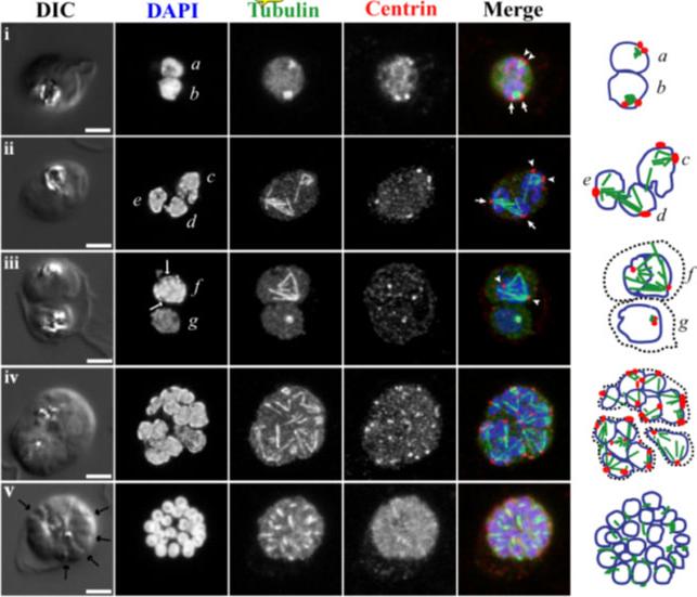
Fluorescence light microscopy images of mitotic 450 spindle during blood stage schizogony in P. falciparum parasites. 3D confocal microscopy views of blood-stage P.f. parasite cell morphology (DIC microscopy), nuclei (DAPI, blue in merged images), mitotic spindle (anti-alpha tubulin antibody, green in merged images), and mitotic spindle MTOCs (anti-PfCEN3 antibody in panels i, ii, iv, and v or anti-Cr Centrin1 antibody 20H5 in panel iii, red in merged images). Schematic cartoons are drawn for each example indicating spindle MTOCs (red circles), microtubules (green lines), and outlines of stained DNA (blue lines). Dotted black lines indicate separate parasites in multiply infected host cells. Panel i, a-b) Short mitotic spindles bounded by MTOCs. The spindle in nucleus b which resembles an oblong tubulin spot has a pole-to-pole distance that is approximately 1 micron long. Panels ii-iii, c-f) Larger spindle structures. In d and e, spindle extends across separate nuclear bodies. In f, a dark line is visible down the center of the DAPI-stained DNA near the spindle midzone (DAPI, arrows). Panel iv) Asynchronous schizont nuclei have spindles with different geometries in multiple stages of extension. Panel v) Segmented parasites after the final mitosis of schizogony. Furrows are visible between daughters (arrows), nuclei are condensed, and microtubules appear in the daughter cytoplasms. Panel i is a single confocal slice from a 3D series. Panels ii-v are maximum projection images of full 3D series. Panels ii-iv were processed with deconvolution software. Scale bar = 2 microns. Gerald N, Mahajan B, Kumar S. Mitosis in the human malaria parasite Plasmodium falciparum. Eukaryot Cell. 2011 10(4):474-82.
See original on MMP
Upper panel: P. falciparum asexual stage schizonts were stained with a mixture of mouse anti-PfCEN3 (Alexa Fluor 488 label) and anti-a-tubulin PFI0180w (direct Alexa Fluor 680 label) antibodies or stained separately. DAPI staining the parasite nucleus is shown in blue channel in panel 1; PfCEN3 localization is shown on the green channel in panel 2; a-tubulin localization is shown on the red channel in panel 3. Merging of the blue, green, and red channels is shown on panel 4. The green dots represent the centrin localization to the poles of hemi-spindles and full spindles, the predicted localization of centrosomes (upper row). The association of centrin alongside of the DAPI-stained nucleus is shown in the lower row. Scale bar, 10 mm. When stained with PfCEN3 antibody alone, centrin reactivity appears as a pattern of paired spots that are organized around DAPI-positive nuclei.Lower panel: Developing rhoptry pairs are arranged in a symmetrical pattern around PfCEN3 centrosomes. Confocal image of a schizont stage parasite. Nuclei are stained with DAPI (blue), anti-PfCEN3 antibody is labeled green, and anti-RAP1 antibody is labeled red to stain rhoptries. Rhoptries (red) are arranged in pairs around the peripheral cytoplasm of the developing schizont (RAP1, arrows). PfCEN3-positive centrosomes (green) are located between each rhoptry pair (Merge, arrows).Mahajan B, Selvapandiyan A, Gerald NJ, Majam V, Zheng H, Wickramarachchi T, Tiwari J, Fujioka H, Moch JK, Kumar N, Aravind L, Nakhasi HL, Kumar S. Centrins, cell cycle regulation proteins in human malaria parasite Plasmodium falciparum. J Biol Chem. 2008 283:31871-83.
See original on MMP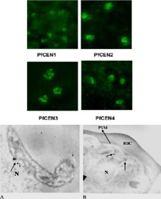
Expression of P. falciparum centrin in schizonts. P. falciparum parasites were probed against mouse anti-PfCEN1, -2, -3, and -4 antibodies. Centrins are associated with the mitotic spindle. Immunoelectron micrograph showing reactivity with the P. falciparum centrin-2 antibody. The gold particles associated with the nucleus of P. falciparum sporozoite stage parasite (A) and nucleus of asexual blood stage schizonts (B). N, nucleus; PVM, parasitophorous vacuole membrane; RBC, red blood cell. In sporozoites, IEM localization of PfCEN2 was observed as multiple dots in an area surrounding the nucleus suggesting a possible association with the centrosome. Similarly, in blood form schizonts, immunogold particles were seen to be associated with an area in proximity to the nucleus.Mahajan B, Selvapandiyan A, Gerald NJ, Majam V, Zheng H, Wickramarachchi T, Tiwari J, Fujioka H, Moch JK, Kumar N, Aravind L, Nakhasi HL, Kumar S. Centrins, cell cycle regulation proteins in human malaria parasite Plasmodium falciparum. J Biol Chem. 2008 283:31871-83.
See original on MMP
e, Nuclear development in PfCRK4-depleted parasites (D10 parent, representative of three biological replicates, and results with P2G12-PfCRK4-HA-DD). Scale bar, 2 μm. f, Immunofluorescence detection of spindle structures in PfCRK4-depleted parasites (P2G12 parent, representative of two biological replicates). Tub, tubulin. Scale bar, 2 μm. g, Quantification of centriolar plaques by immunofluorescence in PfCRK4-depleted parasites (D10 parent; P value, chi-square test; representative of two biological replicates and results with P2G12-PfCRK4-HA-DD). Cen, centrin. Scale bar, 1 μm. Analysis of nuclei stained with fluorescent DNA-specific dyes confirmed that the nuclei did not divide and revealed a substantiallydistorted nuclear morphology (e). Hemispindle structures were evident in PfCRK4-depleted cells (f); however, they were greatlyenlarged relative to spindles in wild-type parasites and might account for the nuclear distortion.Ganter M, Goldberg JM, Dvorin JD, Paulo JA, King JG, Tripathi AK, Paul AS, Yang J, Coppens I, Jiang RH, Elsworth B, Baker DA, Dinglasan RR, Gygi SP, Duraisingh MT. Plasmodium falciparum CRK4 directs continuous rounds of DNA replication during schizogony. Nat Microbiol. 2017 2:17017
See original on MMPMore information
| PlasmoDB | PF3D7_1027700 |
| GeneDB | PF3D7_1027700 |
| Malaria Metabolic Pathways | Localisation images Pathways mapped to |
| Previous ID(s) | PF10_0271, PF3D7_1027700.1, PF3D7_1027700.2 |
| Orthologs | PBANKA_0511800 , PCHAS_0511900 , PKNH_0612100 , PVP01_0612800 , PVX_111335 , PY17X_0512900 |
| Google Scholar | Search for all mentions of this gene |