PF3D7_1022500 citrate synthase, mitochondrial, putative (CS)
Disruptability [+]
| Species | Disruptability | Reference | Submitter |
|---|---|---|---|
| P. falciparum 3D7 |
Possible |
25843709 | Theo Sanderson, Wellcome Trust Sanger Institute |
| P. falciparum 3D7 |
Possible |
USF piggyBac screen (Insert. mut.) | USF PiggyBac Screen |
Mutant phenotypes [+]
| Species | Stage | Phenotype | Reference | Submitter |
|---|---|---|---|---|
| P. falciparum 3D7 | Asexual |
No difference |
25843709 | Theo Sanderson, Wellcome Trust Sanger Institute |
Imaging data (from Malaria Metabolic Pathways)
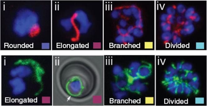
Top - Apicoplast and mitochondrial morphology and association during schizogony in P. falciparum. Live citrate synthase-YFP and acyl carrier proten DsRed double transfectant cells were co-labelled with the nuclear dye Hoechst 33258. Apicoplast morphology was categorized into four sorts: (i) rounded (ii) elongated (iii) branched and (iv) divided. Results indicated that at the onset of schizogony, the apicoplast is predominantly elongated in form. By the 6–10 nucleus stage, branched apicoplasts are the predominant form, with the majority of apicoplasts divided by the >15 nucleus stage. Bottom - Mitochondrial morphology was categorized as (i and ii) elongated (iii) branched and (iv) divided. The mitochondrion is a dynamic organelle, whose predominant form throughout the asexual cycle was branched. Mitochondrial division occurs very late in schizogony. The mitochondrion appears capable of fusion (ii and iii) and frequently associates with the plasma membrane (ii, arrow).van Dooren GG, Marti M, Tonkin CJ, Stimmler LM, Cowman AF, McFadden GI. Development of the endoplasmic reticulum, mitochondrion and apicoplast during the asexual life cycle of Plasmodium falciparum. Mol Microbiol. 2005 57:405-19.
See original on MMP
Apicoplast division precedes mitochondrial division during schizogony. Acyl carrier protein (ACP) is an apicoplast marker. By mid-schizont stage, the majority of mitochondria are branched and the apicoplast also branches (A). Occasionally apicoplast branches appear to associate with mitochondrial branches (A arrows). The branched apicoplast always divides before the mitochondrion (B–D). Each segregated apicoplast closely associates with a branch of the mitochondrion (B–D). Fourth panel from the left in B shows a close-up of the boxed area in the third panel. Rarely, we observe apicoplasts not associated with a mitochondrial branch (arrow in C). The mitochondrion remains branched until apparently quite late in schizogony. Upon division, each mitochondrion associates with a divided apicoplast, and this organellar pair segregates into a single daughter merozoite. Arrow in E shows a mitochondrion associated with two apicoplasts. Presumably, this mitochondrion is still in the process of division.van Dooren GG, Marti M, Tonkin CJ, Stimmler LM, Cowman AF, McFadden GI. Development of the endoplasmic reticulum, mitochondrion and apicoplast during the asexual life cycle of Plasmodium falciparum. Mol Microbiol. 2005 57:405-19.
See original on MMP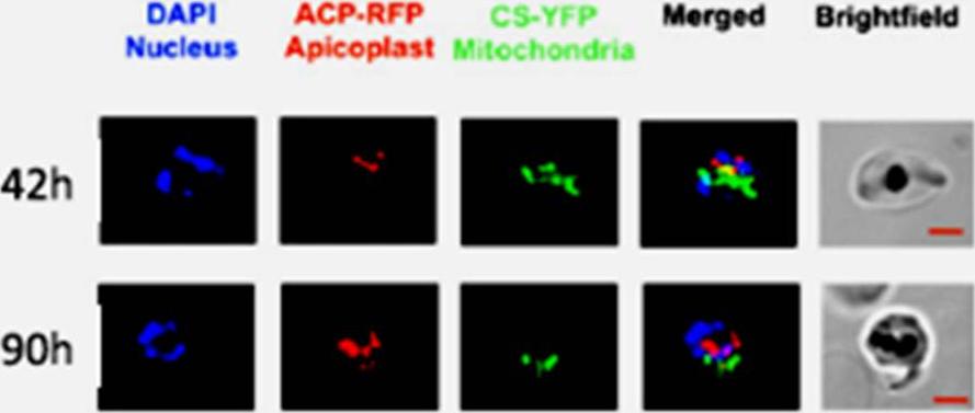
Plasmodium endosymbiotic organelles were visualised using double-transfected parasites with red fluorescent protein-labelled apicoplasts and yellow fluorescent protein-labeled mitochondria (D10 acyl carrier protein-RFP, citrate synthetase-YFP). In the schizont stage both the apicoplast and mitochondria elongate, branch and divide to segregate into daughter merozoites. Jackson KE, Pham JS, Kwek M, De Silva NS, Allen SM, Goodman CD, McFadden GI, Ribas de Pouplana L, Ralph SA. Dual targeting of aminoacyl-tRNA synthetases to the apicoplast and cytosol in Plasmodium falciparum. Int J Parasitol. 2012 42:177-86. PMID: .
See original on MMP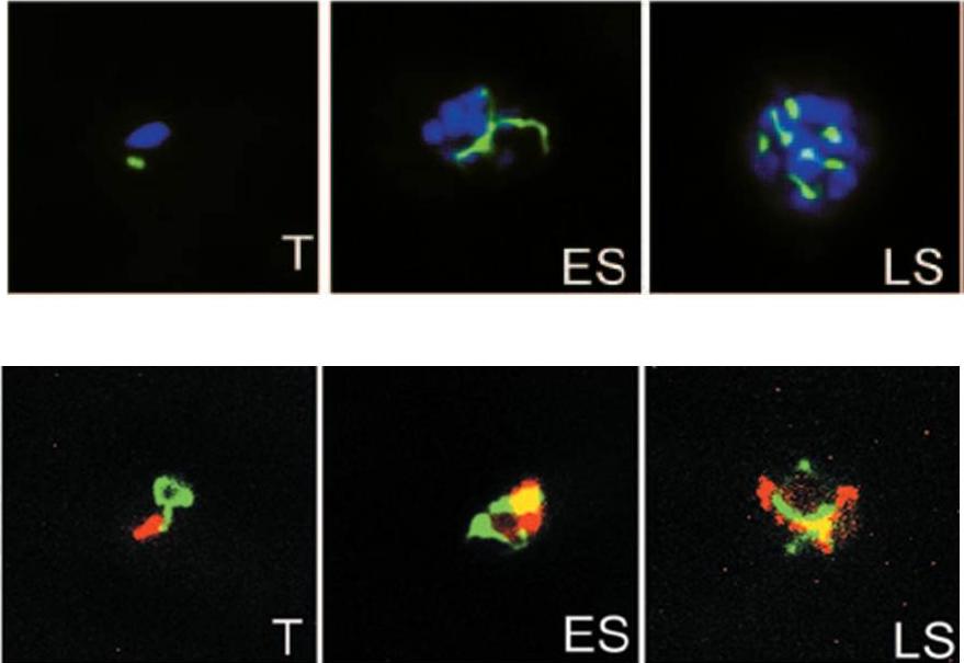
Upper panel: Parasites stably expressing the apicoplast targeted ACPl-GFP transgene were analyzed by fluorescence microscopy at trophozoite (T), early schizont (ES), and late schizont (LS) stages. The apicoplast appears green; nuclei are stained blue with DAPI.Lower panel: Dually transfected parasites expressing the mitochondrion-targeted CSl-YFP transgene and the apicoplast-targeted ACPl-DsRed transgene were analyzed by fluorescence microscopy at trophozoite (T), early schizont (ES), and late schizont (LS) stages are shown. The mitochondrion appears green, and the apicoplast appears red,Dahl EL, Shock JL, Shenai BR, Gut J, DeRisi JL, Rosenthal PJ. Tetracyclines specifically target the apicoplast of the malaria parasite Plasmodium falciparum. Antimicrob Agents Chemother. 2006 50:3124-31. PubMed PMID:
See original on MMP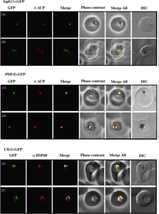
Immuno-localization of GFP chimeras in transgenic parasites using a new Plasmodium falciparum cell fixation protocol. IspE(l)-GFP and PDF(f)-GFP were fixed and incubated with anti-ACP rabbit anti-serum. IspE(l)-GFP (A and B) co-localizes with anti-ACP. PDF(f)-GFP also co-localizes with anti-ACP (C and D), although it sometimes appears to localize within a sub-region of the ACP labelling (D). Stable CS(l)-GFP expressing cells (E and F) were incubated in the presence of anti-Hsp60 from E.coli. Overlay of GFP with Hsp60 suggests this Hsp60 PF10_0153 antibody is a useful mitochondrial marker. Transmission images were captured in both phase contrast and differential interference contrast (DIC) to highlight the integrity of cells fixed under these new conditions.Tonkin CJ, van Dooren GG, Spurck TP, Struck NS, Good RT, Handman E, Cowman AF, McFadden GI. Localization of organellar proteins in Plasmodium falciparum using a novel set of transfection vectors and a new immunofluorescence fixation method. Mol Biochem Parasitol. 2004 137:13-21. Copyright Elsevier 2009.
See original on MMP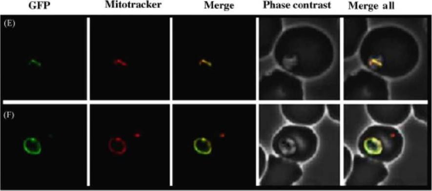
CS(l)-GFP (E and F) fluoresces as a more elongated intracellular structure in early stage parasites, which co-localizes with MitoTracker.Tonkin CJ, van Dooren GG, Spurck TP, Struck NS, Good RT, Handman E, Cowman AF, McFadden GI. Localization of organellar proteins in Plasmodium falciparum using a novel set of transfection vectors and a new immunofluorescence fixation method. Mol Biochem Parasitol. 2004 137:13-21. Copyright Elsevier 2009. PMID:
See original on MMP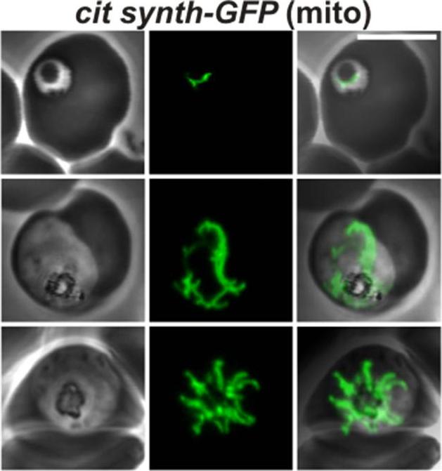
Confocal fluorescence microscopy images of transfected 3D7 P. falciparum-infected RBCs expressing a GFP chimera directed to the mitochondrion. DIC image, the GFP fluorescence signal and an overlay of a P. falciparum citrate synthase-GFP transfectant. Scale bar = 5µm. In ring stage cells, the mitochondrion appears as a single tubular organelle (top row). In the trophozoite stage, the mitochondrion extends to form branched structures (middle row). During schizogony, the mitochondrion divides to provide an organelle for each daughter cell (bottom row). Tilley L, McFadden G, Cowman A, Klonis N. Illuminating Plasmodium falciparum-infected red blood cells. Trends Parasitol. 2007 23:268-77. Copyright Elsevier 2009.
See original on MMP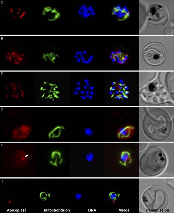
Clindamycin and tetracycline treatment results in “delayed-death” and significant defects in the apicoplast and mitochondrial morphology. Scanning confocal microscopy images of live P. falciparum expressing ACPLRFP (apicoplast - red) and CSLYFP (mitochondria - green) and stained with Hoescht 33342 (DNA—blue) after 48 h without drug treatment. (E) Images taken after 48 h treatment with 100nM clindamycin. (F) Images taken after 48 h treatment with 10mM tetracycline. (G) Images of clindamycin treated parasite after 72 h. (H) Images of tetracycline treated parasite after 72 h. Arrow indicates spherical body which may be the apicoplast. (I) Images of untreated parasite after 72 h.Goodman CD, Su V, McFadden GI. The effects of anti-bacterials on the malaria parasite Plasmodium falciparum. Mol Biochem Parasitol. 2007 52(2):181-91
See original on MMP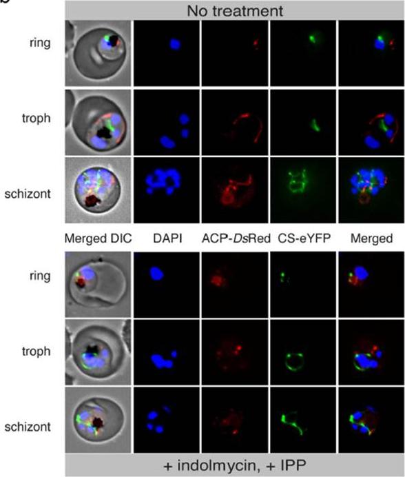
Loss of apicoplast in indolmycin-treated, IPP-rescued parasites. Live cell fluorescence comparing treated and untreated ACP-DsRed/CS-eYFP P. falciparum 3D7. ACP, apicoplast acyl-carrier protein. DIC, differential interference contrast. CS, mitochondrial citrate synthase. Pasaje CF, Cheung V, Kennedy K, Lim EE, Baell JB, Griffin MD, Ralph SA. Selective inhibition of apicoplast tryptophanyl-tRNA synthetase causes delayed death in Plasmodium falciparum. Sci Rep. 2016 6:27531.
See original on MMPMore information
| PlasmoDB | PF3D7_1022500 |
| GeneDB | PF3D7_1022500 |
| Malaria Metabolic Pathways | Localisation images Pathways mapped to |
| Previous ID(s) | PF10_0218 |
| Orthologs | PBANKA_0506700 , PCHAS_0506800 , PKNH_0606800 , PVP01_0607700 , PVX_111595 , PY17X_0507800 |
| Google Scholar | Search for all mentions of this gene |