PF3D7_1021800 schizont egress antigen-1 (SEA1)
Disruptability [+]
| Species | Disruptability | Reference | Submitter |
|---|---|---|---|
| P. falciparum 3D7 |
Refractory |
24855263 | Theo Sanderson, Wellcome Trust Sanger Institute |
| P. falciparum 3D7 |
Possible |
USF piggyBac screen (Insert. mut.) | USF PiggyBac Screen |
Mutant phenotypes [+]
| Species | Stage | Phenotype | Reference | Submitter |
|---|---|---|---|---|
| P. falciparum 3D7 | Asexual |
Egress defect |
24855263 (Conditional) | Theo Sanderson, Wellcome Trust Sanger Institute |
| P. falciparum 3D7 | Asexual |
Refractory |
24855263 (Conditional) | Theo Sanderson, Wellcome Trust Sanger Institute |
Imaging data (from Malaria Metabolic Pathways)
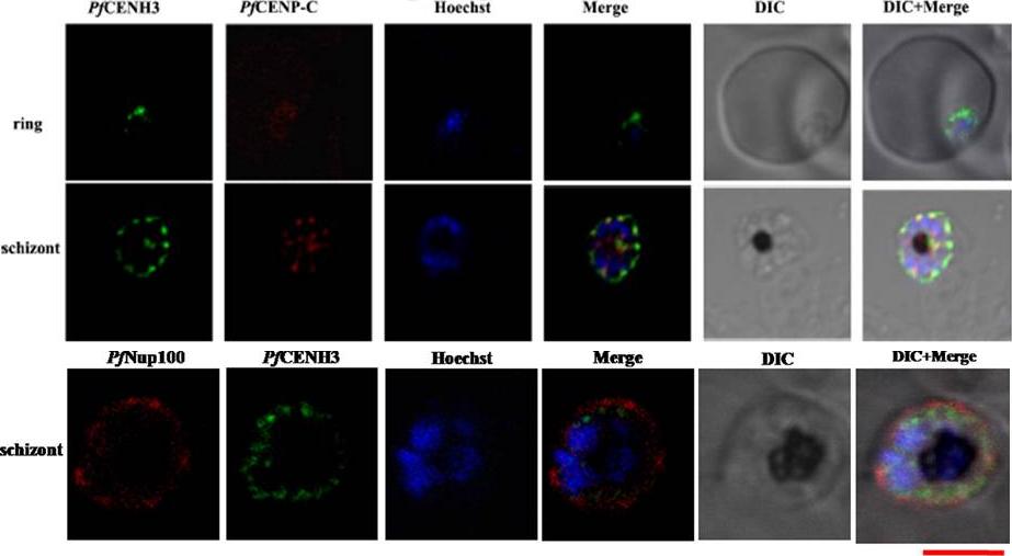
Upper: Colocalization of PfCENH3 with PfCENP-C. Confocal images showing the co-localization of the putative PfCENP-C (red) and PfCENH3 (green) at the nuclear periphery. The nucleus is stained with Hoechst (blue). Scale bar = 5 mm. Lower: The fluorescence pattern obtained on merging PfCENH3 and PfNup100with the nucleus depicts that PfCENH3 localizes to the nuclear periphery just outside the Hoechst stained nucleus but well within the PfNup100 that marks the outer nuclear boundaries.Verma G, Surolia N. Plasmodium falciparum CENH3 is able to functionally complement Cse4p and its, C-terminus is essential for centromere function. Mol Biochem Parasitol. 2013 Dec 6. [Epub ahead of print]
See original on MMP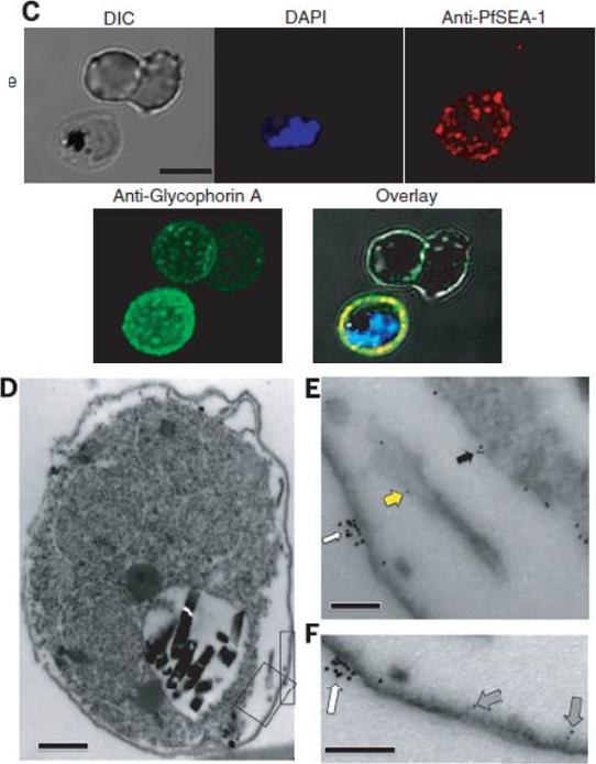
Immunolocalization of PfSEA-1. (C) Nonpermeabilized, unfixed schizont-infected RBCs were probed with mouse anti–PfSEA-1 (red) and rabbit anti–glycophorin A (green) and counterstained with DAPI to label parasite nuclei. PfSEA-1 colocalized with glycophorin A to the surface of schizont-infected RBCs. Scale bar, 5 mm. (D to F) Nonpermeabilized, unfixed schizontinfected RBCs were probed with mouse anti–PfSEA-1 (5-nm gold particles) and rabbit anti–glycophorin A (10-nm gold particles). PfSEA-1 localized to the schizont/parasitophorous vacuole membrane (black arrow), Maurer's clefts (yellow arrow), and the inner leaflet of the RBC membrane (gray arrow), whereas glycophorin A was confined to the outer leaflet of the RBC membrane (white arrow). Scale bar in (D), 0.5 mm. Scale bars in (E) and (F), 0.1 mm.Raj DK, Nixon CP, Nixon CE, Dvorin JD, DiPetrillo CG, Pond-Tor S, Wu HW, Jolly G, Pischel L, Lu A, Michelow IC, Cheng L, Conteh S, McDonald EA, Absalon S, Holte SE, Friedman JF, Fried M, Duffy PE, Kurtis JD. Antibodies to PfSEA-1 block parasite egress from RBCs and protect against malaria infection. Science. 2014 344(6186):871-7.
See original on MMP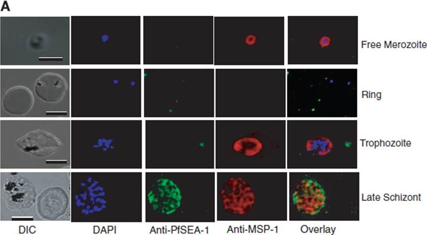
Immunolocalization of PfSEA-1. (A) Infected RBCs were probed with mouse anti–PfSEA-1 (green) and rabbit anti–MSP-1 (red) and counterstained with 4',6'-diamidino-2-phenylindole (DAPI) to label parasite nuclei. PfSEA-1 is detected only in schizont-infected RBCs. Scale bar for free merozoite images, 2 mm. Scale bar for remaining images, 10 mm. Anti–PfSEA-1 did not bind to free merozoites, rings, or late trophozoite–stage parasites, but did specifically recognize an antigen expressed by late schizont–infected RBCs.Raj DK, Nixon CP, Nixon CE, Dvorin JD, DiPetrillo CG, Pond-Tor S, Wu HW, Jolly G, Pischel L, Lu A, Michelow IC, Cheng L, Conteh S, McDonald EA, Absalon S, Holte SE, Friedman JF, Fried M, Duffy PE, Kurtis JD. Antibodies to PfSEA-1 block parasite egress from RBCs and protect against malaria infection. Science. 2014 344(6186):871-7.
See original on MMP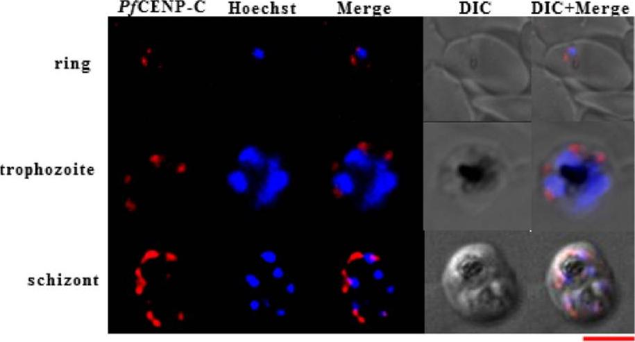
PfCENP-C exhibits constitutive differential expression throughout the intraerythrocytic stages and localize to the nuclear periphery. The confocal images for immunofluorescence assays representing the subcellular localization pattern of PfCENP-C (red) and the Hoechst stained nucleus (blue) at ring, trophozoite and schizont stages of P. falciparum, respectively. Scale bar = 5 μm. Verma G, Surolia N. The dimerization domain of PfCENP-C is required for its functions as a centromere protein in human malaria parasite Plasmodium falciparum. Malar J. 2014 Dec 4;13(1):475. PMID:
See original on MMP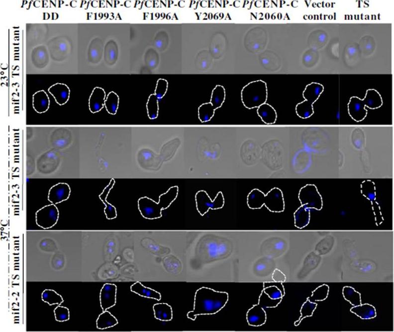
Functionally essential residues lie within the PfCENP-C dimerization domain. the phenotypic changes exhibited by the dimerization domain mutants PfCENP-CF1993A, PfCENPCF1996A, PfCENP-CY2069A and PfCENP-CN2060A were assessed at 23 and 37°C, respectively. At 23°C, mif2-3 and mif2-2 TS mutant cells expressing different PfCENP-C dimerization domains show proper chromosome segregation and normal bud morphology. At 37°C, ~90-94% of mif2-3 and mif2-2 TS mutant cells expressing PfCENP-CF1993A, PfCENP-CF1996A and PfCENP-CY2069A are mitotically arrested and unviable. This is evident by the aberrant bud morphologies with fragmented nucleus or loss of nuclear staining. These abnormal phenotypes are attributable to the failure of PfCENP-C dimerization domain mutants to dimerize resulting in impaired growth, defective chromosome segregation and loss of viability. Verma G, Surolia N. The dimerization domain of PfCENP-C is required for its functions as a centromere protein in human malaria parasite Plasmodium falciparum. Malar J. 2014 Dec 4;13(1):475.
See original on MMP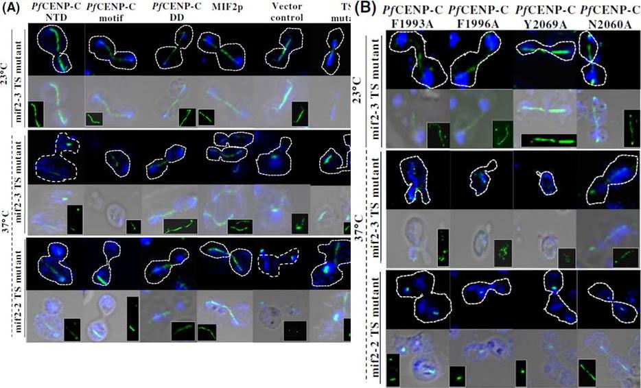
The mitotic spindle integrity is maintained by the PfCENP-C dimerization domain. (A) Confocal images showing the mitotic spindle (green) and the Hoechst stained nucleus (blue) of the mif2-3 and mif2-2 TS mutant cells expressing the PfCENP-C-NTD, -motif and -DD, region, MIF2p and empty vector alone at 23 and 37°C, respectively. At 23°C, all the mif2-3 TS mutants expressing different constructs show normal spindle structure connecting the nuclear masses across the bud and mother cell. At 37°C, both in mif2-3 and mif2-2 TS mutants except the PfCENP-C-DD and MIF2p, all the other constructs show abnormal and discontinuous spindles. The spindle morphology (green) is also shown in the insets. The broken spindle in themif2-3 and mif2-2 TS mutant cells are shown by red arrows in the insets. B) The confocal images representing the disintegrated mitotic spindle morphologies in mif2-3 and mif2-2 TS mutant cells expressing the PfCENP-C-DD mutants, PfCENP-CF1993A, PfCENP-CF1996A and PfCENP-CY2069A while PfCENP-CN2060A show normal mitotic spindles at 37°C. NTD- N-terminal region; DD-dimerization domain.Verma G, Surolia N. The dimerization domain of PfCENP-C is required for its functions as a centromere protein in human malaria parasite Plasmodium falciparum. Malar J. 2014 Dec 4;13(1):475.
See original on MMPMore information
| PlasmoDB | PF3D7_1021800 |
| GeneDB | PF3D7_1021800 |
| Malaria Metabolic Pathways | Localisation images Pathways mapped to |
| Previous ID(s) | PF10_0212, PF10_0212a |
| Orthologs | PBANKA_0506000 , PCHAS_0506100 , PKNH_0606100 , PVP01_0607000 , PVX_001955 , PY17X_0507100 |
| Google Scholar | Search for all mentions of this gene |