PF3D7_1017300 golgi re-assembly stacking protein 1, golgi re-assembly stacking protein 2 (GRASP)
Disruptability [+]
| Species | Disruptability | Reference | Submitter |
|---|---|---|---|
| P. falciparum 3D7 |
Possible |
USF piggyBac screen (Insert. mut.) | USF PiggyBac Screen |
Mutant phenotypes [+]
None reported yet. Please press the '+' button above to add one.Imaging data (from Malaria Metabolic Pathways)
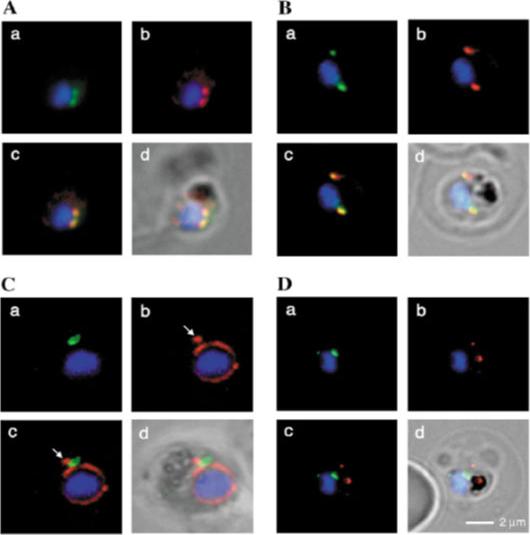
Fluorescence microscopy on fixed parasites. (A) PfGRASP-GFP colocalises with antiPfGRASP-specific antibodies. PfGRASP-GFP is tightly confined to two compartments (a, green) near the parasite nucleus (a, blue). AntiPfGRASP-specific antibodies show a similar staining pattern (b, red with nucleus in blue). Merged image shows identical colocalisation. (B) PfGRASP-GFP colocalises with the cis-Golgi marker ERD2. PfGRASP-GFP (a, green) accumulates in two discrete compartments in close proximity to the nucleus (a, blue). Anti-PfERD2 antibodies recognize similar structures (b, red with nucleus in blue). Merged image shows colocalisation of compartments (c, yellow). (C) PfGRASP-GFP defines a compartment that is distinct from the ER. At the early stages of the parasite life cycle (<16 hours post invasion) PfGRASP is restricted to one compartment (a, green) juxtapose to the nucleus (a, blue). The ER is visualised by anti-PfBiP-specific antibodies (b, red). The membranous system of the ER forms an envelope around the nucleus (b, blue) with one protrusion (indicated by arrow). Merged image shows no colocalisation of the two compartments (c). (D) PfGRASP-GFP does not colocalise with the trans-Golgi marker PfRab6. PfGRASP accumulates in two discrete foci (a, green) adjacent to the nucleus (a, blue). Antibodies against PfRab6 visualise two distinct sites within the parasite (b, red with nucleus in blue). Merged image shows no colocalisation of the PfGRASP defined compartment with PfRab6 (c).Struck NS, de Souza Dias S, Langer C, Marti M, Pearce JA, Cowman AF, Gilberger TW. Re-defining the Golgi complex in Plasmodium falciparum using the novel Golgi marker PfGRASP. J Cell Sci. 2005 118(Pt 23):5603-13.
See original on MMP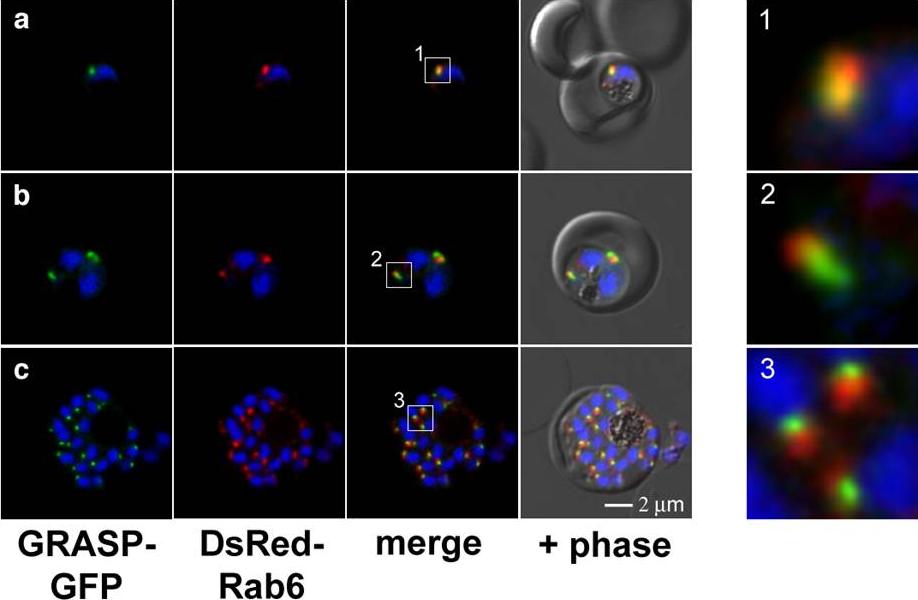
Cis- versus trans-Golgi dynamics during the asexual life cycle of P. falciparum. GRASP–GFP and DsRed–Rab6 were used to visualize the spatial organization of the cis- and trans face of the Golgi in ring stages (a), early (b) and late schizonts (c). The initially close connection between the cis- and trans-face of the Golgi in young parasites (a) appears to expand during maturation (b), resulting in a clearly distinct trans-Golgi compartment in schizonts (c). The boxed regions in a-c are depicted in higher magnification in 1-3.Struck NS, Herrmann S, Schmuck-Barkmann I, de Souza Dias S, Haase S, Cabrera AL, Treeck M, Bruns C, Langer C, Cowman AF, Marti M, Spielmann T, Gilberger TW. Spatial dissection of the cis- and trans-Golgi compartments in the malaria parasite Plasmodium falciparum. Mol Microbiol. 2008 67:1320-30.
See original on MMP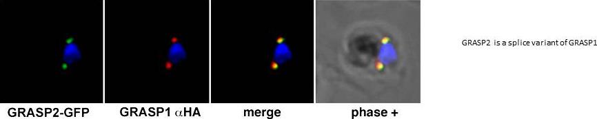
Colocalization of GRASP 1 and 2 in a double transgenic parasite line (Golgi re-assembly stacking protein). Immunofluorescence assay of GRASP2-GFP (green) with HA-specific antibodies representing GRASP1 (red). The merged image shows colocalisation of the two compartments. All images show the nucleus in blue (DAPI).Reproduced with permission from Struck et al., 2008: Plasmodium falciparum possesses two GRASP proteins that are differentially targeted to the Golgi complex via a higher- and lower-eukaryote-like mechanism. J. Cell Sci. 121: 2123-29.
See original on MMP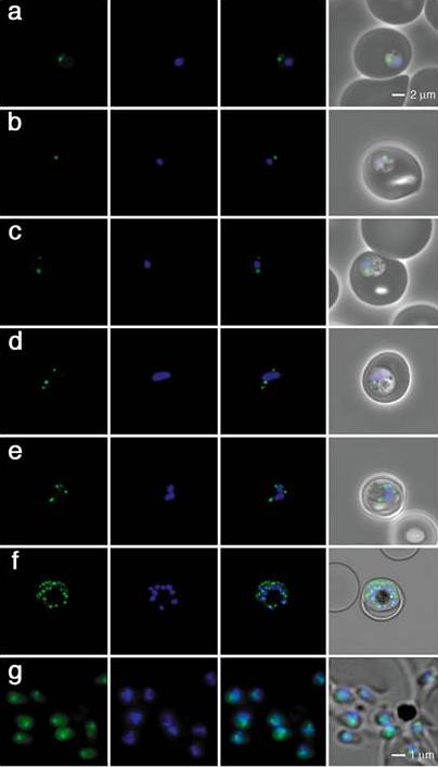
Live images of transgenic parasites expressing PfGRASP-GFP were visualized by confocal microscopy. In ring-stage parasites (8-16 hours post invasion, a-b) PfGRASP-GFP is restricted to one compartment in close proximity to the nucleus (blue). A second Golgi is generated prior to nuclear division (24 hours post invasion, c). As the parasite matures, nuclear division commences and is accompanied by multiplication of the Golgi (32-40 hours post invasion, d-e). The parasite has nearly reached the final stage of schizogony where each forming merozoite will be equipped with one Golgi and nucleus (46 hours post invasion, f). Each merozoite has inherited one Golgi (0 hours, g).Reproduced with permission from Struck et al., 2005: Re-defining the Golgi complex in P. falciparum using the novel Golgi marker PfGRASP. J. Cell Sci. 118: 5603-13.
See original on MMP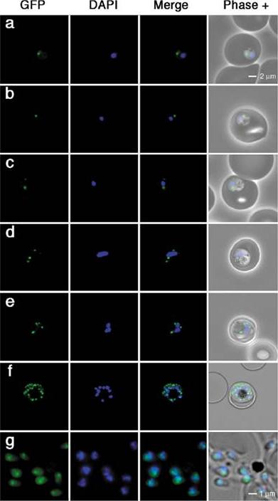
Golgi dynamics throughout the asexual life cycle of Plasmodium. Live images of transgenic parasites expressing PfGRASP-GFP were visualised by confocal microscopy. (a-b) Images 8-16 hours post invasion. In ring-stage parasites PfGRASP-GFP is restricted to one compartment in close proximity to the nucleus (blue). (c) Images 24 hours post invasion. A second Golgi is generated prior to nuclear division. (d-e) Cells 32-40 hours post invasion. As the parasite matures, nuclear division commences and is accompanied by multiplication of the Golgi. (f) Cells 46 hours post invasion. The parasite has nearly reached the final stage of schizogony where each forming merozoite will be equipped with one Golgi and nucleus. (g) Released parasites at 0 hours. Each merozoite has inherited one Golgi. Bar, 2 m (a-f); 1 mm (g).Struck NS, de Souza Dias S, Langer C, Marti M, Pearce JA, Cowman AF, Gilberger TW. Re-defining the Golgi complex in Plasmodium falciparum using the novel Golgi marker PfGRASP. J Cell Sci. 2005 118(Pt 23):5603-13.
See original on MMP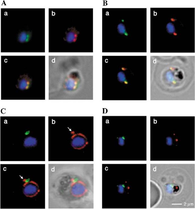
Spatial organisation of the PfGRASP-GFP defined compartment by fluorescence microscopy on fixed parasites. (A) PfGRASP-GFP colocalises with anti PfGRASP-specific antibodies. PfGRASP-GFP is tightly confined to two compartments (a, green) near theparasite nucleus (a, blue). AntiPfGRASP-specificantibodies show a similar staining pattern (b, red withnucleus in blue). Merged image shows the colocalisation of the compartments defined by either PfGRASP-specific antibodies or PfGRASP-GFP expressingparasites (c, yellow). (B) PfGRASP-GFP colocalises with the cis-Golgi marker ERD2. PfGRASP-GFP (a, green) accumulates in two discrete compartments in close proximity to the nucleus (a, blue). Anti-PfERD2 antibodies recognize similar structures (b, red with nucleus in blue). Merged image shows colocalisation of compartments (c, yellow). (C) PfGRASP-GFP defines a compartment that is distinct from the ER . At the early stages of the parasite life cycle (<16 hours post invasion) PfGRASP is restricted to one compartment (a, green) juxtapose to the nucleus (a, blue). The ER is visualised by anti-PfBiP-specific antibodies (b, red). The membranous system of the ER forms an envelope around the nucleus (b, blue) with one protrusion (indicated by arrow). Merged image shows no colocalisation of the two compartments (c). (D) PfGRASP-GFP does not colocalise with the trans-Golgi marker PfRab6. PfGRASP accumulates in two discrete foci (a, green) adjacent to the nucleus (a, blue). Antibodies against PfRab6 visualise two distinct sites within the parasite (b, red with nucleus in blue). Merged image shows no colocalisation of the PfGRASP defined compartment with PfRab6 (c) All panels labelled d in A-D are merges of fluorescent and bright-field images. Bar, 2 mm. Struck NS, de Souza Dias S, Langer C, Marti M, Pearce JA, Cowman AF, Gilberger TW. Re-defining the Golgi complex in Plasmodium falciparum using the novel Golgi marker PfGRASP. J Cell Sci. 2005 118(Pt 23):5603-13.
See original on MMP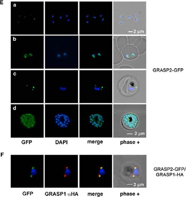
(E) Localisation of GRASP2-GFP in unfixed parasites. Free merozoites display GFP fluorescence in one tightly defined compartment (a). A duplication of this compartment takes place in ring-stage parasites prior to nuclear division (b). As the parasite matures from trophozoite (c) to schizont (d) the organelle further multiplies until one compartment can be equally distributed among the forming progeny. (F) Colocalisation of the two GRASP proteins in the double transgenic parasite line. Immunofluorescence assay of GRASP2-GFP (green) with HA-specific antibodies representing GRASP1 (red). The merged image shows colocalisation of the two compartments. One single GRASP2-GFP compartment in close proximity to the nucleus was observed. in free merozoites (Ea). Duplication of the GRASP2-defined compartment occured prior to nuclear division (Eb) resulting in a multiplicity of Golgi compartments in schizonts (Ec,d).Struck NS, Herrmann S, Langer C, Krueger A, Foth BJ, Engelberg K, Cabrera AL, Haase S, Treeck M, Marti M, Cowman AF, Spielmann T, Gilberger TW. Plasmodium falciparum possesses two GRASP proteins that are differentially targeted to the Golgi complex via a higher- and lower-eukaryote-like mechanism. J Cell Sci. 2008 121(Pt 13):2123-9.
See original on MMP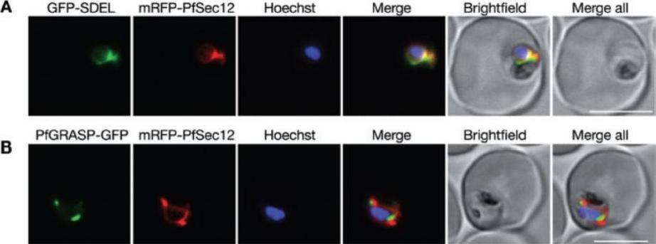
Comparison of PfSec12 localization with markers for the ER and Golgi. Parasites were cotransfected with plasmids for expression of mRFP-PfSec12 (from the native pfsec12 5′ UTR) with either (A) GFP-SDEL, a marker for the general ER, or (B) PfGRASP-GFP, a cis-Golgi marker. mRFP-PfSec12 was localized throughout the ER coincident with GFP-SDEL, in close apposition to PfGRASP-labelled Golgi. Bar = 5 μm.Lee MC, Moura PA, Miller EA, Fidock DA. Plasmodium falciparum Sec24 marks transitional ER that exports a model cargo via a diacidic motif. Mol Microbiol. 2008 68(6):1535-46.
See original on MMP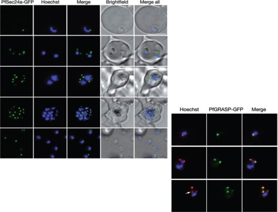
Left Panel: Live cell imaging of parasites expressing PfSec24-GFP (PF13_0324), with nuclei stained with Hoechst 33342. PfSec24a marks distinct foci that likely represent transitional ER sites that are enriched in the COPII vesicle machinery. Parasites at the ring (row 1), trophozoite (row 2), early schizont (row 3) and late schizont (row 4) stages, as well as released merozoites (row 5) are shown. Bar = 5 µM. Right panel: Close apposition of PfSec24a-marked tER sites and Golgi. Parasites expressing PfSec24a–mRFP and PfGRASP–GFP demonstrate a close relationship between the number and location of tER sites and Golgi, as illustrated in three representative parasites.Lee MC, Moura PA, Miller EA, Fidock DA. Plasmodium falciparum Sec24 marks transitional ER that exports a model cargo via a diacidic motif. Mol Microbiol. 2008 68:1535-46.
See original on MMP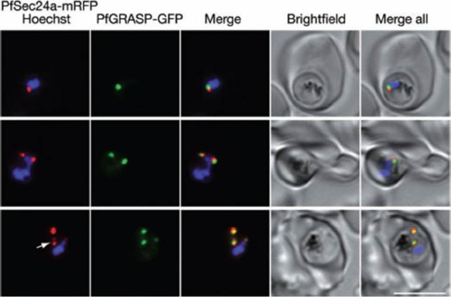
Close apposition of PfSec24a-marked tER sites and Golgi. Parasites expressing PfSec24a-mRFP and PfGRASP-GFP demonstrate a close relationship between the number and location of tER sites and Golgi, as illustrated in three representative parasites. Occasionally, two tER foci can be observed adjacent to a single Golgi spot (arrow in bottom row). Bar = 5 μm.Lee MC, Moura PA, Miller EA, Fidock DA. Plasmodium falciparum Sec24 marks transitional ER that exports a model cargo via a diacidic motif. Mol Microbiol. 2008 68(6):1535-46.
See original on MMP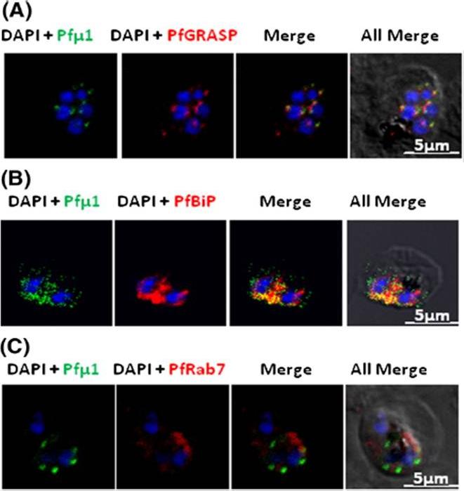
Pfμ1 is closely associated with the Golgi and ER in early trophozoite stages. Transgenic parasites expressing Pfμ1–GFP protein at trophozoite stages were immunostainedwith anti-PfGRASP (A), anti-PfBiP (B) and anti-PfRab7 (C) antibodies. The parasite nuclei were stained with DAPI and slides were visualized by confocal laser scanning microscopy. Scale bars denote 5 μm. Pfμ1 partially co-localized with PfGRASP and PfBip. the ER (which is contiguous with the nuclear envelope) and Golgi in developing merozoites are very closely juxtaposed, and dense vesicular traffic occurs between these two compartments, as well as between nascent rhoptries. No overlap between Pfμ1–GFP and Rab7.This suggests that these proteins do not co-localize, and hence that Rab7 is not involved in trafficking at this stage (3C). These results thus suggest the presence of Pfμ1 in the Golgi–ER network during the early stages of intra-erythrocytic development.Kaderi Kibria KM, Rawat K, Klinger CM, Datta G, Panchal M, Singh S, Iyer GR, Kaur I, Sharma V, Dacks JB, Mohmmed A, Malhotra P. A role for adaptor protein complex 1 in protein targeting to rhoptry organelles in Plasmodium falciparum. Biochim Biophys Acta. 2015 1853(3):699-710.
See original on MMP
GFP-Rab18 fluorescence co-localizes with ER/Golgi, dense granules or microneme markers. GFP-PfRab1A fluorescence is distinct from the localization of markers for the apicoplast (ACP), the ER (Bip), the Golgi (ERD2 and GRASP), as well as from staining of the ER/Golgi with Bodipy BFA.Morse D, Webster W, Kalanon M, Langsley G, McFadden GI. Plasmodium falciparum Rab1A Localizes to Rhoptries in Schizonts. PLoS One. 2016 11(6):e0158174
See original on MMP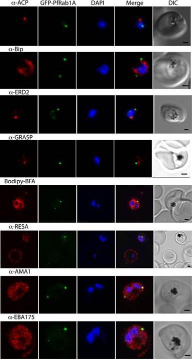
GFP-PfRab1A fluorescence does not co-localize with ER/Golgi, dense granules or microneme markers. GFP-PfRab1A fluorescence is distinct from the localization of markers for the apicoplast (ACP), the ER (Bip), the Golgi (ERD2 and GRASP), as well as from staining of the ER/Golgi with Bodipy BFA. GFP-Rab1A fluorescence is also distinct from the localization of markers for dense granules (RESA) or micronemes (AMA1 and EBA175).Morse D, Webster W, Kalanon M, Langsley G, McFadden GI. Plasmodium falciparum Rab1A Localizes to Rhoptries in Schizonts. PLoS One. 2016 Jun 27;11(6):e0158174
See original on MMP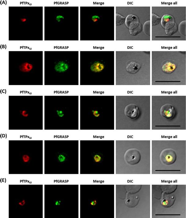
Immunofluorescence images showing the Golgi morphology in AlF4 and vinblastine treated parasites. (A) PfGRASP localization in control parasites for AlF4treatment, (B) Golgi morphology in AlF4- treated parasites (17 parasites counted, Golgi structure was dispersed in 95% parasites), (C) Pf-GRASP localization in solvent (PBS) control parasites for vinblastine treatment, (D) Golgi morphology in vinblastine-treated parasites (18 parasites counted, Golgi structure was dispersed in 95% parasites),(E) Golgi morphology in parasites reverted after vinblastine treatment (11 parasites counted, Intact Golgi structure was observed in 90% parasites). Scale Bar: 10 mm.Chaudhari R, Dey V, Narayan A, Sharma S, Patankar S. Membrane and luminal proteins reach the apicoplast by different trafficking pathways in the malaria parasite Plasmodium falciparum. PeerJ. 2017 Apr 27;5:e3128.
See original on MMPMore information
| PlasmoDB | PF3D7_1017300 |
| GeneDB | PF3D7_1017300 |
| Malaria Metabolic Pathways | Localisation images Pathways mapped to |
| Previous ID(s) | PF10_0168, PF3D7_1017300.1, PF3D7_1017300.2 |
| Orthologs | PBANKA_0501600 , PCHAS_0501700 , PKNH_0601500 , PVP01_0602500 , PVX_001735 , PY17X_0502600 |
| Google Scholar | Search for all mentions of this gene |