PF3D7_1015900 enolase (ENO)
Disruptability [+]
| Species | Disruptability | Reference | Submitter | |
|---|---|---|---|---|
| P. falciparum 3D7 |
Refractory |
USF piggyBac screen (Insert. mut.) | USF PiggyBac Screen | |
| P. berghei ANKA |
Refractory |
PlasmoGEM (Barseq) | PlasmoGEM | |
Mutant phenotypes [+]
None reported yet. Please press the '+' button above to add one.Imaging data (from Malaria Metabolic Pathways)
Immunofluorescence images showing variation in distribution of enolase between cytosol and nucleus in a population of synchronized ring stage parasite cells. There were greater number of cells having more enolase signal arising from nucleus than from cytosol.Pal Bhowmick I, Kumar N, Sharma S, Coppens I, Jarori GK. Plasmodium falciparum enolase: stage-specific expression and sub-cellular localization. Malar J. 2009 8(1):179.
See original on MMP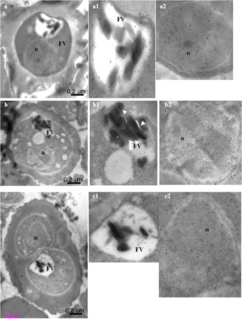
Figure 4. Immuno-gold electron microscopic (IEM) imaging for the localization of enolase in early trophozoite satge of P. falciparum using mouse anti-r-Pfen antibody. Magnified views of the food vacuole (FV) and nucleus (n) are also shown. Arrows in food vacuole marks hemozoin associated enolase. Enolase was found to be associated with cytosol,nucleus, food vacuole, cytoskeleton and plasma membrane.Pal Bhowmick I, Kumar N, Sharma S, Coppens I, Jarori GK. Plasmodium falciparum enolase: stage-specific expression and sub-cellular localization. Malar J. 2009 8(1):179. PubMed
See original on MMP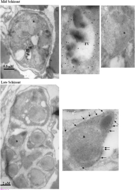
Immuno-gold electron microscopic (IEM) imaging for the localization of enolase in mid and late stage schizonts of P. falciparum using mouse anti-r-Pfen antibody. Magnified views of the food vacuole (FV) and nucleus (n) are also shown. Presence of enolase on the surface of a merozoite is marked with arrows. Enolase was found to be associated with cytosol,nucleus, food vacuole, cytoskeleton and plasma membrane.Pal Bhowmick I, Kumar N, Sharma S, Coppens I, Jarori GK. Plasmodium falciparum enolase: stage-specific expression and sub-cellular localization. Malar J. 2009 8(1):179. PubMed
See original on MMP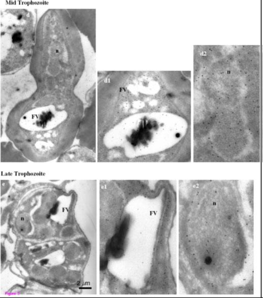
Immuno-gold electron microscopic (IEM) imaging for the localization of enolase in mid and late stage trophozoites of P. falciparum using mouse anti-r-Pfen antibody. Magnified views of the food vacuole (FV) and nucleus (n) are also shown. Enolase was found to be associated with cytosol,nucleus, food vacuole, cytoskeleton and plasma membrane.Pal Bhowmick I, Kumar N, Sharma S, Coppens I, Jarori GK. Plasmodium falciparum enolase: stage-specific expression and sub-cellular localization. Malar J. 2009 8(1):179. PubMed
See original on MMP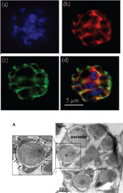
IFA of the schizont stage of P. falciparum using DAPI (a), rabbit anti-r-Pfen antiserum (1:200) with secondary anti-rabbit IgG conjugated with AlexaFluor 568 (b), and mouse anti-MSP-1 antibodies (1:100) with secondary anti-mouse IgG conjugated with AlexaFluor 488 (c). An overlay of the images from frames a, b, and c is shown in frame d. The enolase protein is localized on the merozoite cell surface.A. IEM image using mouse anti-r-Pfen antiserum at a 1:100 dilution with a P. falciparum-infected red cell at young trophozoite stage the presence of enolase on the parasite plasma membrane (ppm) is marked with arrows. Hpm, host cell plasma membrane; n, nucleus.Pal-Bhowmick I, Mehta M, Coppens I, Sharma S, Jarori GK. Protective properties and surface localization of Plasmodium falciparum enolase. Infect Immun. 2007 75:5500-8.
See original on MMP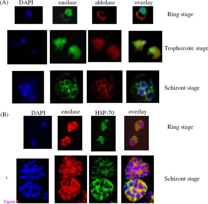
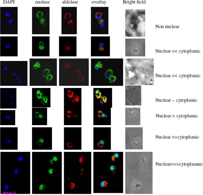
Immunofluorescence assays for the localization of enolase, aldolase and HSP-70 in P. falciparum asexual stages (ring, trophozoite and schizont). (A) P. falciparum infected red blood cells were treated with DAPI (blue), mouse anti rPfen antibody (green), rabbit anti-P. falciparum aldolase antibody (red). (B) Cells were treated with DAPI, rabbit rPfen antibody (red), and mouse anti Pf HSP-70 antibody (green). Overlay panels show the merged of the three images.Pal Bhowmick I, Kumar N, Sharma S, Coppens I, Jarori GK. Plasmodium falciparum enolase: stage-specific expression and sub-cellular localization. Malar J. 2009 8(1):179
See original on MMP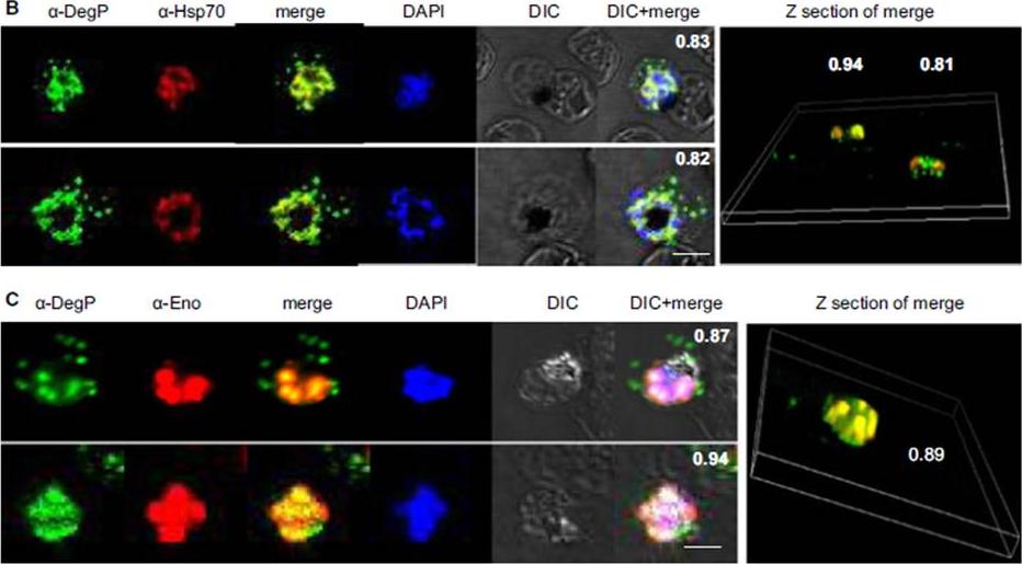
PfDegP exists in a multimeric complex, and is associated with parasite proteins Hsp70 and Eno. Parasites were stained with anti-DegP and anti-Hsp70 (B) or anti-Eno (C) antiserum; parasite nuclei were stained with 4′,6-diamidino-2-phenylindole (DAPI). Fluorescence signals from DegP and Hsp70 or Eno antibodies showed significant merging, with a co-localization coefficient > 0.80.Sharma S, Jadli M, Singh A, Arora K, Malhotra P. A secretory multifunctional serine protease, DegP of Plasmodium falciparum, plays an important role in thermo-oxidative stress, parasite growth and development. FEBS J. 2014 Mar;281(6):1679-99.
See original on MMPMore information
| PlasmoDB | PF3D7_1015900 |
| GeneDB | PF3D7_1015900 |
| Malaria Metabolic Pathways | Localisation images Pathways mapped to |
| Previous ID(s) | PF10_0155 |
| Orthologs | PBANKA_1214300 , PCHAS_1215000 , PKNH_0816200 , PVP01_0816000 , PVX_095015 , PY17X_1217500 |
| Google Scholar | Search for all mentions of this gene |