PF3D7_1015200 cysteine--tRNA ligase, putative (CysRS)
Disruptability [+]
| Species | Disruptability | Reference | Submitter |
|---|---|---|---|
| P. falciparum 3D7 |
Refractory |
USF piggyBac screen (Insert. mut.) | USF PiggyBac Screen |
Mutant phenotypes [+]
None reported yet. Please press the '+' button above to add one.Imaging data (from Malaria Metabolic Pathways)

Live-cell epifluorescence microscopy of P. falciparum transfected using pGlux.1 vector for episomal expression of GFP fused to the two Pf CysRS mRNA isoforms: Pf CysRS-A1–153GFP (A) and Pf CysRS-B1–70GFP (B) in live cells. In (A), both parasites are early trophozoite stage, and localization of GFP is to a single punctum, consistent with the size and position of the apicoplast. The red blood cell (RBC) and the parasite (P) are indicated. (B) An early trophozoite (top panels) and late trophozoite (bottom panels) with GFP dispersed throughout the cytosol. (C) Immunofluorescence assays of the parasites indicated, Pf CysRS-A1–153GFP (top panels) and Pf CysRS-B1–70GFP (bottom panels), using antibodies against GFP and ACP, an apicoplast marker. The Pf CysRS-A1–153GFP signal specifically co-localizes with the apicoplast marker, whereas the Pf CysRS-B1–70GFP signal is distributed throughout the cytosol. For (A)–(C), all scale bars indicate 5 μm. Pham JS, Sakaguchi R, Yeoh LM, De Silva NS, McFadden GI, Hou YM, Ralph SA. A dual-targeted aminoacyl-tRNA synthetase in Plasmodium falciparum charges cytosolic and apicoplast tRNACys. Biochem J. 2014 458(3):513-23
See original on MMP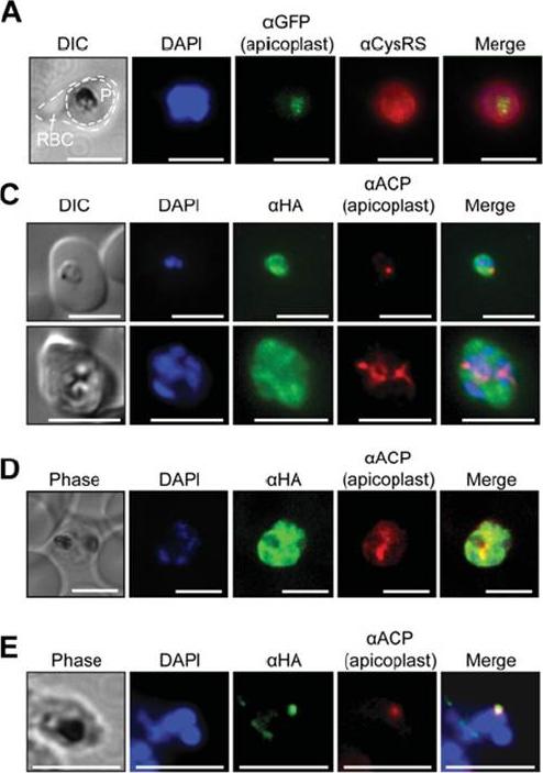
(A) Immunofluorescence assays of trophozoite stage P. falciparum (3D7 strain) using an anti-Pf CysRS antibody, DAPI and the apicoplast marker PfRRf1–GFP. The red blood cell is indicated (RBC) and the parasite is indicated (P). (C) Epifluoresence microscopy and (D) confocal imaging of immunofluorescence analysis of Pf CysRS–HA transfectants at early- and late-trophozoite stage parasites (top and bottom panels respectively) with an anti-HA antibody against the Pf CysRS–HA enzyme, ananti-ACP antibody for the apicoplast, and DAPI for the nucleus. The images show that the Pf CysRS–HA is distributed throughout the cytosol, and also overlap with the apicoplast marker ACP. The width of the apicoplast is less than the spatial resolution of light microscopy, sowe cannot definitively assign co-localization from these experiments. (D) Confocal images of Pf CysRS–HA transfectant parasites at late-trophozoite stage using indirect immunofluorescence analysis reveals cytoplasmic distribution ofPf CysRS–HA overlapping with the apicoplast marker ACP. (E) Immunofluorescence analysis with saponin-treated Pf CysRS–HA to differentially lysemembranes to allow for visualizing subcellular organelles with antibodies and stains as indicated. Parasites are labelled with an anti-HA antibody against the Pf CysRS–HA enzyme, and an anti-ACP antibody as a marker of the apicoplast. This immunolocalization confirms that afraction of Pf CysRS is specifically retained within the apicoplast. All scale bars indicate 5 μm.Pham JS, Sakaguchi R, Yeoh LM, De Silva NS, McFadden GI, Hou YM, Ralph SA. A dual-targeted aminoacyl-tRNA synthetase in Plasmodium falciparum charges cytosolic and apicoplast tRNACys. Biochem J. 2014 458(3):513-23
See original on MMPMore information
| PlasmoDB | PF3D7_1015200 |
| GeneDB | PF3D7_1015200 |
| Malaria Metabolic Pathways | Localisation images Pathways mapped to |
| Previous ID(s) | PF10_0149, PF3D7_1015200.1, PF3D7_1015200.2, PF3D7_1015200.3 |
| Orthologs | PBANKA_1213600 , PCHAS_1214300 , PKNH_0815300 , PVP01_0815300 , PVX_094980 , PY17X_1216800 |
| Google Scholar | Search for all mentions of this gene |