PF3D7_1012200 rhoptry associated adhesin (RA)
Disruptability [+]
| Species | Disruptability | Reference | Submitter | |
|---|---|---|---|---|
| P. falciparum 3D7 |
Refractory |
USF piggyBac screen (Insert. mut.) | USF PiggyBac Screen | |
| P. berghei ANKA |
Possible |
PlasmoGEM (Barseq) | PlasmoGEM | |
Mutant phenotypes [+]
| Species | Stage | Phenotype | Reference | Submitter |
|---|---|---|---|---|
| P. berghei ANKA | Asexual |
No difference |
PlasmoGEM (Barseq) | PlasmoGEM |
Imaging data (from Malaria Metabolic Pathways)

Native protein expression of PfRA in P. falciparum. Immunofluorescence assays were performed on fixed smears of different stages of the P. falciparum intraerythrocytic life cycle. PfRA (green) staining was detected by the anti-PfRA mouse antibodies only in the schizont stages. No signal was observed in trophozoites and schizonts. A distinct punctate staining of PfRA was observed in the schizont which is characteristic of apically localized proteins. Pre-immune antibodies failed to detect any staining. Nuclei were stained with the DNA intercalating dye, DAPI. (Scale bar, 2 μm.)Anand G, Reddy KS, Pandey AK, Mian SY, Singh H, Mittal SA, Amlabu E, Bassat Q, Mayor A, Chauhan VS, Gaur D. A novel Plasmodium falciparum rhoptry associated adhesin mediates erythrocyte invasion through the sialic-acid dependent pathway. Sci Rep. 2016 6:29185.
See original on MMP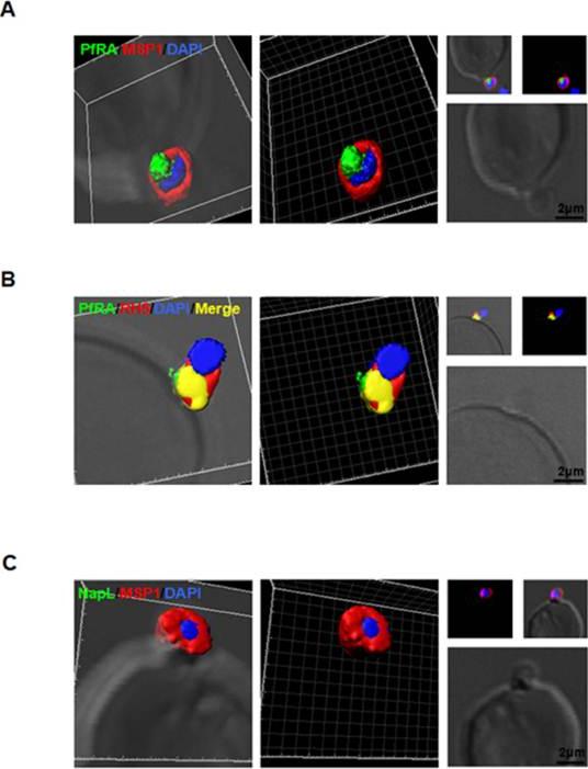
PfRA is present at the apical surface of an invading merozoite. Confocal immunofluorescencemicroscopy analysis of PfRA localization in cytochalasin-D treated invading merozoites under non-permeable fixing conditions using glutaraldehyde /paraformaldehyde. (A) 3D reconstruction of z-stacks during merozoite invasion co-immunostained with MSP-1and PfRA. PfRA detected at the apical end of the merozoite surface. In the 3D images, the γ settings were altered for visual representation only. (B) PfRA (green) co-localizes with rhoptry bulb marker PfRH5 (red) that is known to be translocated to the merozoite surface to engage with its erythrocyte receptor, Basigin. (C) As a control, staining of a nuclear parasite protein, NapL, was not detected under the same non-permeable fixing conditions confirming that the fluorescent signals were obtained only from surface localized proteins. Nuclei were stained with DNA intercalating dye DAPI (Scale bar, 2 μ m.)Anand G, Reddy KS, Pandey AK, Mian SY, Singh H, Mittal SA, Amlabu E, Bassat Q, Mayor A, Chauhan VS, Gaur D. A novel Plasmodium falciparum rhoptry associated adhesin mediates erythrocyte invasion through the sialic-acid dependent pathway. Sci Rep. 2016 6:29185.
See original on MMP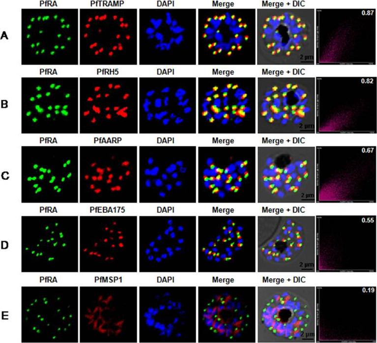
PfRA localizes to the rhoptries of P. falciparum schizonts and merozoites. Co-localization of PfRA with (A) PfTRAMP, (B) PfRH5, (C) PfAARP, (D) PfEBA-175 and (E) MSP-1 in schizont stage parasites by confocal immunoflourescence microscopy. PfRA (green) co-localizes with rhoptry bulb protein PfTRAMP(red) and PfRH5 (red) but does not co-localize with the micronemal protein PfEBA-175 (red) and merozoite surface protein MSP-1 in schizonts. PfRA (green) showed partial co-localization with rhoptry neck protein PfAARP (red). The co-localization of PfRA was quantitatively analyzed and the Pearson correlation co-efficientwith PfTRAMP (0.87) and PfRH5 (0.82) suggested that the two proteins were co-localized with PfRA. The coefficients of PfRA with PfAARP, PfEBA-175 and MSP1 were lower than 0.7 indicating that the three proteins were not co-localized with PfRA. Nuclei were stained with DNA intercalating dye DAPI. (Scale bar, 2 μm.)Anand G, Reddy KS, Pandey AK, Mian SY, Singh H, Mittal SA, Amlabu E, Bassat Q, Mayor A, Chauhan VS, Gaur D. A novel Plasmodium falciparum rhoptry associated adhesin mediates erythrocyte invasion through the sialic-acid dependent pathway. Sci Rep. 2016 6:29185.
See original on MMPMore information
| PlasmoDB | PF3D7_1012200 |
| GeneDB | PF3D7_1012200 |
| Malaria Metabolic Pathways | Localisation images Pathways mapped to |
| Previous ID(s) | PF10_0119 |
| Orthologs | PBANKA_1210600 , PCHAS_1211300 , PKNH_0812000 , PVP01_0812300 , PVX_094830 , PY17X_1213800 |
| Google Scholar | Search for all mentions of this gene |