PF3D7_1008700 tubulin beta chain
Disruptability [+]
| Species | Disruptability | Reference | Submitter | |
|---|---|---|---|---|
| P. berghei ANKA |
Refractory |
PlasmoGEM (Barseq) | PlasmoGEM | |
Mutant phenotypes [+]
None reported yet. Please press the '+' button above to add one.Imaging data (from Malaria Metabolic Pathways)
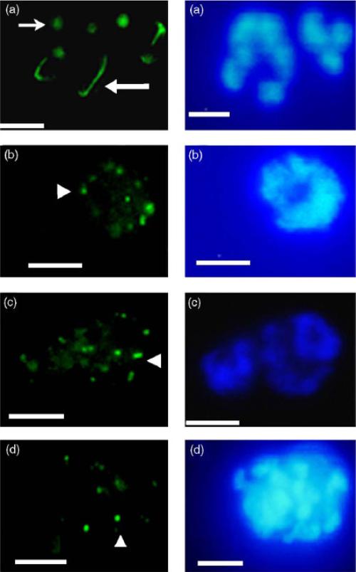
Mitotic microtubular structures of cultured parasites viewed by immunofluorescence using antibodies to P. falciparum b-tubulin (visualised with FITC, left) and DAPI nuclear stain (right). (a) Untreated: note microtubule-organising centres (small arrow) and hemispindles (large arrow); (b) treated with 20mM trifluralin: note dots of tubulin fluorescence (arrowheads) and loss of normal structures; (c) treated with 20mM oryzalin; (d) treated with 20mM APM. In all cases, exposure to inhibitor was for 6 h. Scale bars are 4 mm.Fennell BJ, Naughton JA, Dempsey E, Bell A. Cellular and molecular actions of dinitroaniline and phosphorothioamidate herbicides on Plasmodium falciparum: tubulin as a specific antimalarial target. Mol Biochem Parasitol. 2006 145:226-38. Copyright Elsevier 2011.
See original on MMP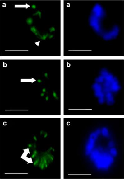
Mitotic and post-mitotic microtubular structures of cultured parasites viewed by immunofluorescence using: (a) anti-P. falciparum b-tubulin antibodies; and (b and c) GT335 (monoclonal anti-polyglutamylated tubulin) visualised with fluorescein isothiocyanate, left) and nuclear bodies stained with DAPI (right). Note microtubule-organising centres (large arrows), hemispindles (arrow head) and radial microtubule spokes (double-arrow). Scale bars =4 mm. Glutamylated tubulins appeared to be limited to the microtubule-organising centres ( b) with no labelling of parasite hemispindles observed. Interestingly, glutamylated tubulins were predominantly located in post-mitotic microtubular structures in a regular radial array, like the spokes of a cartwheel.Fennell BJ, Al-shatr ZA, Bell A. Isotype expression, post-translational modification and stage-dependent production of tubulins in erythrocytic Plasmodium falciparum. Int J Parasitol. 2008 38:527-39. Copyright Elevier 2011.
See original on MMP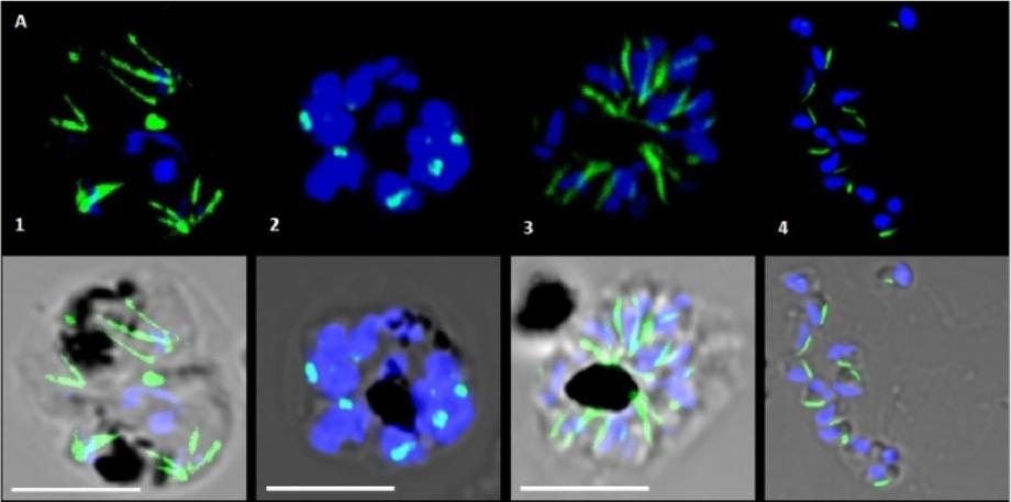
Microtubular structures of P. falciparum as observed in different timelines of progression through the mature blood stages. Parasite tubulin is stained with an anti b tubulin antibody and Alexa Fluor 488 and is shown in green. Blue represents nuclear material stained with DAPI. Panel A: Spindle structures were observed in the mid to late trophozoite stages (A1) and distinct microtubule organizing centers in the schizonts (A2). The typical subpellicular microtubules were seen in the merozoites (A3 and A4). Chakrabarti R, Rawat PS, Cooke BM, Coppel RL, Patankar S. Cellular Effects of Curcumin on Plasmodium falciparum Include Disruption of Microtubules. PLoS One. 2013;8(3):e57302.
See original on MMP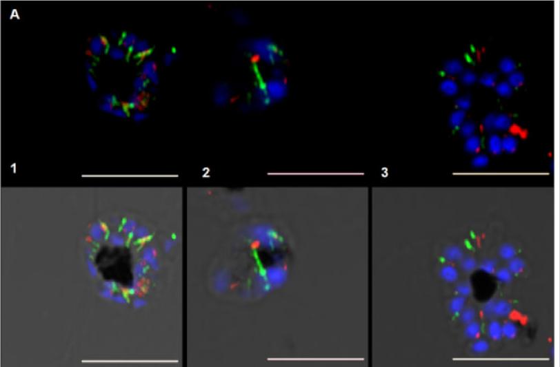
Effect of curcumin on P. falciparum apicoplast. Tubulin fluorescence is depicted in green, apicoplast fluorescence in red and nucleus in blue. Distinct spherical apicoplasts, associated with subpellicular microtubules (A1, A3) and spindle microtubules (A2), were observed. microtubules are known to provide the tracks for segregation of organelles.Chakrabarti R, Rawat PS, Cooke BM, Coppel RL, Patankar S. Cellular Effects of Curcumin on Plasmodium falciparum Include Disruption of Microtubules. PLoS One. 2013;8(3):e57302.
See original on MMP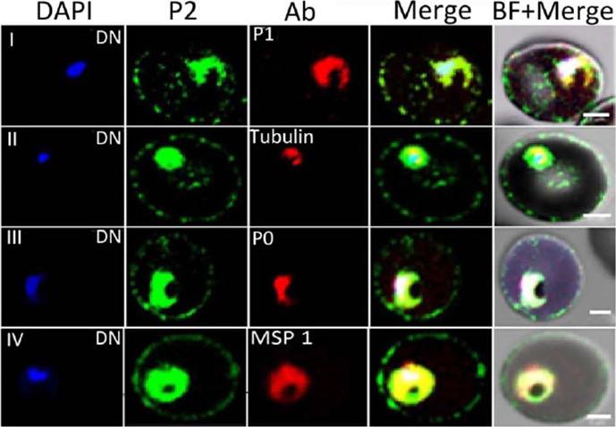
Immunofluorescence assay of P. falciparum infected erythrocytes (IE) using various antibodies. Localization of DAPI (blue), P2 (green); various antibodies (anti-PfP1, anti b-tubulin, anti-PfP0, and anti-MSP1) in red at the di-nuclear (DN) stage of Plasmodium falciparum infected erythrocytes. Scale bar indicates 2 mm.Das S, Basu H, Korde R, Tewari R, Sharma S. Arrest of Nuclear Division in Plasmodium through Blockage of Erythrocyte Surface Exposed Ribosomal Protein P2. PLoS Pathog. 2012 Aug;8(8):e1002858.
See original on MMPMore information
| PlasmoDB | PF3D7_1008700 |
| GeneDB | PF3D7_1008700 |
| Malaria Metabolic Pathways | Localisation images Pathways mapped to |
| Previous ID(s) | PF10_0084 |
| Orthologs | PBANKA_1206900 , PCHAS_1207600 , PKNH_0807700 , PVP01_0808400 , PVX_094635 , PY17X_1210100 |
| Google Scholar | Search for all mentions of this gene |