PF3D7_0936800 Plasmodium exported protein (PHISTc), unknown function
Disruptability [+]
| Species | Disruptability | Reference | Submitter |
|---|---|---|---|
| P. falciparum 3D7 |
Refractory |
18614010 | Theo Sanderson, Wellcome Trust Sanger Institute |
| P. falciparum 3D7 |
Possible |
USF piggyBac screen (Insert. mut.) | USF PiggyBac Screen |
Mutant phenotypes [+]
None reported yet. Please press the '+' button above to add one.Imaging data (from Malaria Metabolic Pathways)
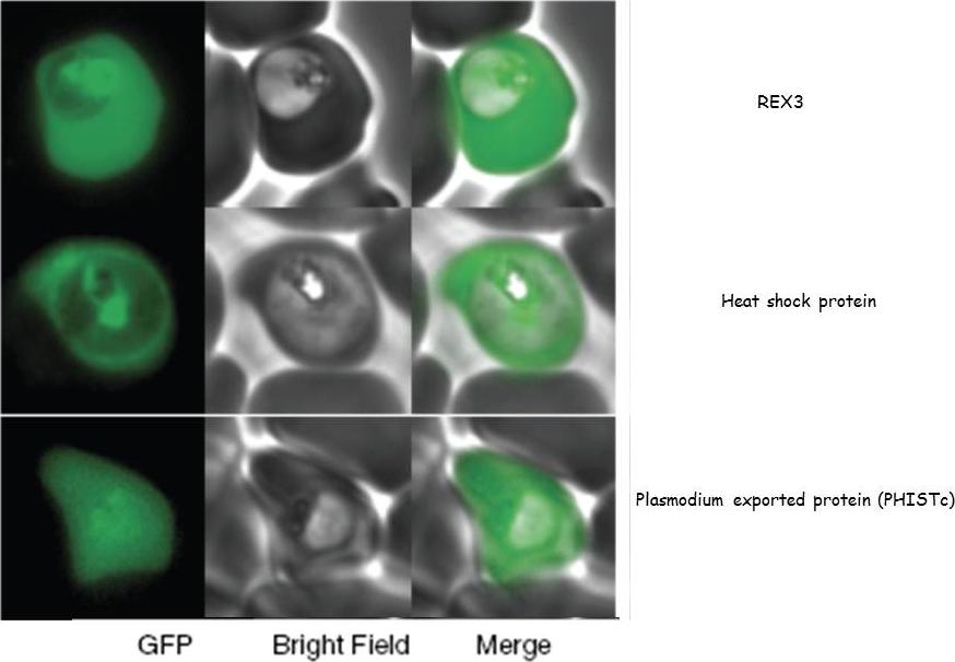
GFP fusions to three genes (PFI1780w, PFE0055c, PFI1755c) whose products are exported into the red blood cell cytosol.Sargeant TJ, Marti M, Caler E, Carlton JM, Simpson K, Speed TP, Cowman AF. Lineage-specific expansion of proteins exported to erythrocytes in malaria parasites. Genome Biol. 2006;7(2):R12;
See original on MMP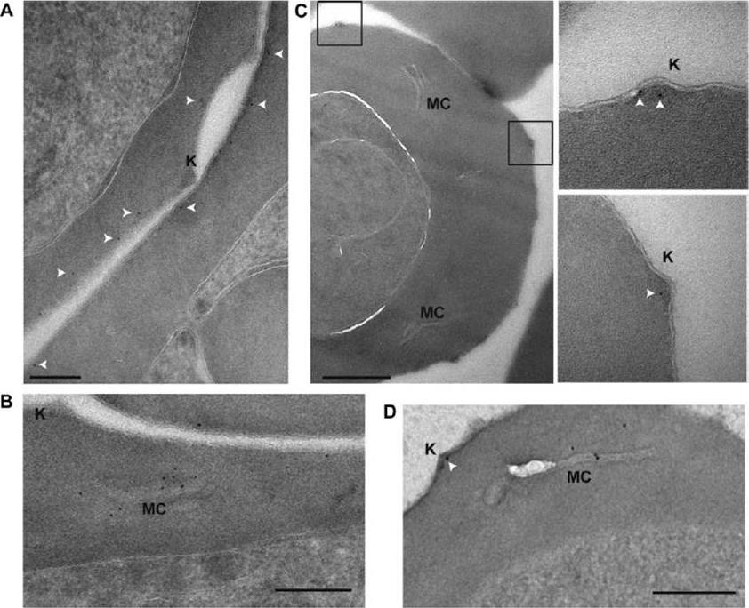
PFE1605w localizes to knobs. Shown here are postembedding immunoelectron microscopy images of iRBCs expressing PFI1780w-GFP (A, B), PFE1605w-HA (C), and PFE1605w-GFP (D). Insets: enlarged views of boxed sections. White arrows, 5 nM gold. Knobs (K) and Maurer’s clefts (MC) are labeled. Scale bars = 200 nm (A); 500 nm (C); 250 nm (B, D). Gold labeling of PFI1780w-GFP was found in proximity to the iRBC membrane but was clearly absent from knobs (A) and frequently at Maurer’s clefts (B). In contrast, PFE1605w-3xHA (C) and PFE1605w-GFP were clearly localized in knobs, and in trophozoite stage parasites, PFE1605w-GFP was also frequently found in Maurer’s clefts (D), suggesting that it localizes underneath the iRBC membrane, whereas PFE1605w localizesto knobs, but both proteins seem to be transiently transported through the Maurer’s clefts.Oberli A, Slater LM, Cutts E, Brand F, Mundwiler-Pachlatko E, Rusch S, Masik MF, Erat MC, Beck HP, Vakonakis I. A Plasmodium falciparum PHIST protein binds the virulence factor PfEMP1 and comigrates to knobs on the host cell surface. FASEB J. 2014 [Epub ahead of print] PMID:
See original on MMP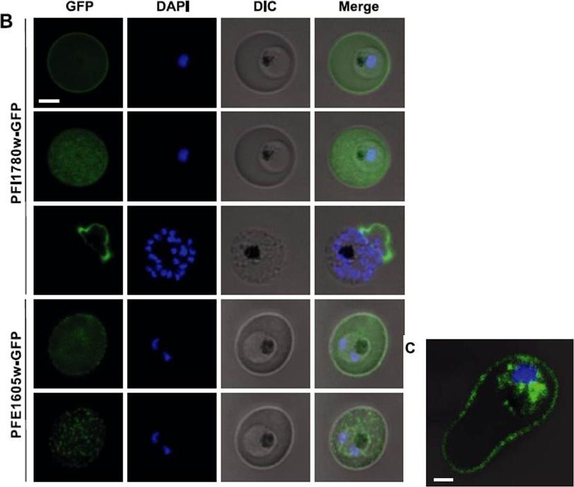
Live cell imaging of 3D7 parasites expressing PFI1780w-GFP and PFE1605w-GFP. For panels 2 and 5, the focal plane of the GFP signal was set on the surface of the iRBC, and the DAPI/differential interference contrast (DIC) microscopy signal was kept on the previous focal plane. Nuclei were stained with DAPI. Scale bar=2 mm. Panel 3 shows a rare schizont with already ruptured erythrocyte membrane. C) Confocal immunofluorescence analysis of 3D7 parasite expressing PFI1780w-GFP. Merge image of GFP/DAPI/DIC channels representing stack 32 of 69 in total. Scale bar =1mm. PFI1780w-GFP was exported to the iRBC cytosol but full-lengthPFI1780w-GFP additionally revealed fluorescence at the periphery of iRBCs (Fig. 2B; top panel), suggesting a localization close or adjacent to the erythrocyte membrane (panel 2). Immunofluorescent 3-dimensional reconstructions of fixed iRBCs with PFI1780w-GFPexpressing parasites showed focal fluorescence in parasite cytosol and uniform luorescence around the biconcave rim of the iRBC (C).Oberli A, Slater LM, Cutts E, Brand F, Mundwiler-Pachlatko E, Rusch S, Masik MF, Erat MC, Beck HP, Vakonakis I. A Plasmodium falciparum PHIST protein binds the virulence factor PfEMP1 and comigrates to knobs on the host cell surface.FASEB J. 2014 [Epub ahead of print]
See original on MMP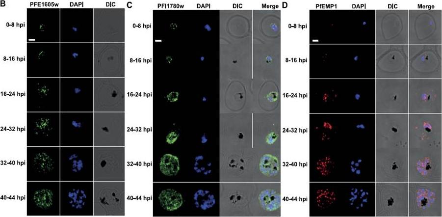
PFE1605w is cotranslocated with PfEMP1. Confocal immunofluorescence analysis of tightly synchronized 3D7 parasites. Samples were collected at 8-h intervals and stained with antibodies recognizing PFE1605w (B), PFI1780w (C), or PfEMP1 (D) and costained with DAPI. Scale bars = 2 mm. DIC, differential interference contrast microscopy. PfEMP1 is known to display a necklace of beads pattern at the parasite surface at ~8–11 hpi which was confirmed (D) and was similar to the pattern observed for PFE1605w (B). Both proteins transiently associated with Maurer’s clefts at ~16–24 hpi (D) before being transferred to the iRBC membrane, suggesting cotransport of PFE1605w with PfEMP1. In contrast to PFE1605w, PFI1780w was found within the parasite cytosol until ~24–32 hpi with limited focal fluorescence in the iRBC cytosol (C). In schizonts, PFI1780w was exported to the iRBC surface as shown in live cell imaging (B) with no distinct intermediate locations.Oberli A, Slater LM, Cutts E, Brand F, Mundwiler-Pachlatko E, Rusch S, Masik MF, Erat MC, Beck HP, Vakonakis I. A Plasmodium falciparum PHIST protein binds the virulence factor PfEMP1 and comigrates to knobs on the host cell surface. FASEB J. 2014 [Epub ahead of print]
See original on MMP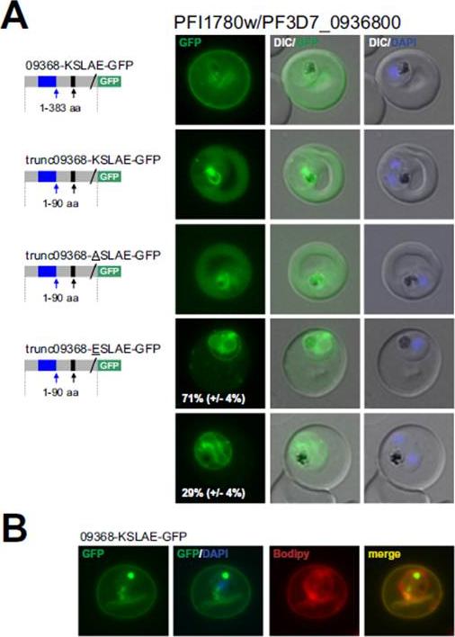
PFI1780w/PF3D7_0936800 encodes an exported PHISTc protein that is cleaved at the non-canonical PEXEL/HT. A. Fluorescence microscopy. The schematic structure of the GFP-tagged proteins is depicted on the left of representative images of iRBCs. The ER-signal is shaded blue, the (non-) canonical PEXEL/HT is shaded black and GFP is shaded green. Putative cleavage sites are indicated by an arrow (blue: SP cleavage site; black: Plasmepsin V cleavage site. For trunc09368-ESLAE-GFP two panels are shown representing iRBCs with reduced export and no export of the reporter protein (at least 50 iRBCs were analyzed on four occasions, whereas ratio is indicated in %; standard deviation in brackets). B. Bodipy-TR-C5-ceramid stained iRBCs expressing 09368_KSLAE-GFP. Fluorescence was located close to the RBC membraneSchulze J, Kwiatkowski M, Borner J, Schlüter H, Bruchhaus I, Burmester T, Spielmann T, Pick C. The Plasmodium falciparum exportome contains non-canonical PEXEL/HT proteins. Mol Microbiol. 2015 Apr 8. [Epub ahead of print]
See original on MMPMore information
| PlasmoDB | PF3D7_0936800 |
| GeneDB | PF3D7_0936800 |
| Malaria Metabolic Pathways | Localisation images Pathways mapped to |
| Previous ID(s) | PFI1780w |
| Orthologs | |
| Google Scholar | Search for all mentions of this gene |