PF3D7_0936300 ring-exported protein 3 (REX3)
Disruptability [+]
| Species | Disruptability | Reference | Submitter |
|---|---|---|---|
| P. falciparum 3D7 |
Possible |
18614010 | Theo Sanderson, Wellcome Trust Sanger Institute |
| P. falciparum 3D7 |
Possible |
USF piggyBac screen (Insert. mut.) | USF PiggyBac Screen |
Mutant phenotypes [+]
None reported yet. Please press the '+' button above to add one.Imaging data (from Malaria Metabolic Pathways)

Upper row: Location of REX3 in IRBCs fixed with 4% formaldehyde/0.005% glutaraldehyde. IFAs with 3D7 parasites expressing a myc-tagged ETRAMP4 showed that REX3 (red; Texas Red) occurs as a uniform stain outside of the PVM (ETRAMP4myc in the PVM detected by a rabbit anti-myc antibody; green, Cy2). Second row: REX3 (red; Texas Red) did not colocalize with the Maurer’s cleft protein REX1 (green; Cy2) in 3D7 parasites. Bars, 5 mm.Lower panel: Confocal fluorescence microscopy images of transfected 3D7 P. falciparum-infected RBCs. REX3 is expressed in its early ring stages (top row) and is directed to the bulk compartment of the host cell cytoplasm. Trophozoites and schizonts (middle and bottom rows) display a uniform fluorescence pattern with some puncta. Tilley L, McFadden G, Cowman A, Klonis N. Illuminating Plasmodium falciparum-infected red blood cells. Trends Parasitol. 2007 23:268-77.
See original on MMP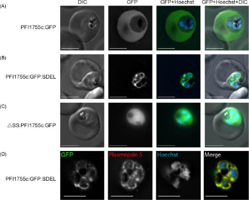
Localization of GFP chimeras in transfected parasites. (A) Localization of PFI1755c:GFP. GFP fluorescence, in live cells, is seen in the erythrocyte cytoplasm indicating that the protein is efficiently exported. (B) Localization of PFI1755c:GFP:SDEL. GFP fluorescence, in live cells, remains within the parasite and shows a characteristic ER distribution. (C) Localization of DSS:PFI1755c:GFP. GFP fluorescence, in live cells, is localized diffusely throughout the cytoplasm of the parasite. Panels show a DIC image, GFP fluorescence, GFP (green) and Hoechst (blue) fluorescence overlaid, and a merge of DIC, GFP (green) and Hoechst (blue) images, as indicated. The scale bar is 4mm. (D) Colocalization of PFI1755c:GFP:SDEL with the ER marker, plasmepsin 5. The GFP fluorescence in PFI1755c:GFP:SDEL expressing parasites was directly visualized and the localization of plasmepsin 5 was determined by indirect immunofluorescence, using an antibody raised against plasmepsin 5 . DNA was stained with Hoechst. In the merged image, GFP fluorescence is in green, plasmepsin 5 staining in red, and regions of co-localization in yellow. The scale bar is 4mm.Osborne AR, Speicher KD, Tamez PA, Bhattacharjee S, Speicher DW, Haldar K. The host targeting motif in exported Plasmodium proteins is cleaved in the parasite endoplasmic reticulum. Mol Biochem Parasitol. 2010 171:25-31. Copyright Elsevier 2010
See original on MMP
(c) A parasite of a line exporting GFP into the host cell and stained with Bodipy-TR-C5-ceramide (red) indicates that the cavity contains host cell cytosol. A single confocal z-section is shown. Note that the nucleus (blue) and the FV (filled with reinternalized GFP) are clearly distinct from the cavity. (d) A cell line exporting GFP into the PV shows a fluorescence pattern similar to Bodipy-TR-C5-ceramide staining (red). A single confocal section is shown. Parasites expressing GFP in the host cell cytosol (using the REX3 export motif) showed fluorescence in the cavity (c), whereas a cell line expressing GFP in the PV (using the EXP1 signal peptide) showed staining similar to Bodipy-TR-C5-ceramide (d), indicating that the cavity represents an invagination of the PVM and the parasite plasma membrane, forming a space filled with host cell cytosol.Grüring C, Heiber A, Kruse F, Ungefehr J, Gilberger TW, Spielmann T. Development and host cell modifications of Plasmodium falciparum blood stages in four dimensions. Nat Commun. 2011 2:165.
See original on MMP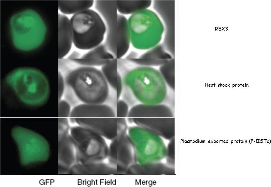
GFP fusions to three genes (PFI1780w, PFE0055c, PFI1755c) whose products are exported into the red blood cell cytosol.Sargeant TJ, Marti M, Caler E, Carlton JM, Simpson K, Speed TP, Cowman AF. Lineage-specific expansion of proteins exported to erythrocytes in malaria parasites. Genome Biol. 2006;7(2):R12;
See original on MMP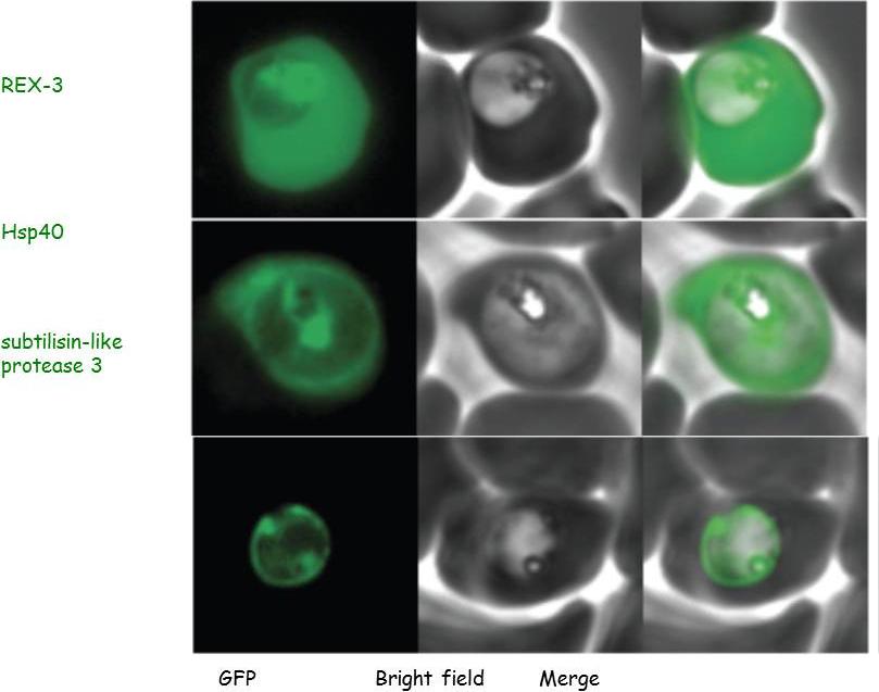
Parasites were transfected with the GFP chimeras of the 3 genes. Experimental verification of their sub-cellular localization indicated that REX-3 and Hsp40 were exported into the host cell cytosol while the protease accumulated in the parasitophorous vacuole.Sargeant TJ, Marti M, Caler E, Carlton JM, Simpson K, Speed TP, Cowman AF. Lineage-specific expansion of proteins exported to erythrocytes in malaria parasites. Genome Biol. 2006; 7(2):R12.
See original on MMP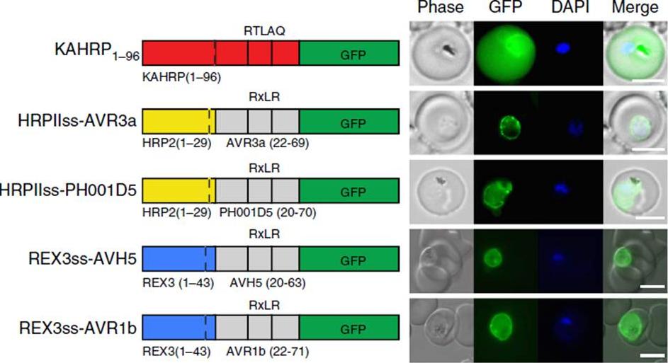
Oomycete RxLR does not mediate export in P. falciparum. (a) KAHRP1–96 is exported to the P. falciparum-infected erythrocyte; however, HRPIIss-AVR3a, HRPIIss-PH001D5, REX3ss-AVH5 and REX3ss-AVR1b are not exported but accumulate in the parasite and PV. Scale bar, 5 mm.Boddey JA, O'Neill MT, Lopaticki S, Carvalho TG, Hodder AN, Nebl T, Wawra S, van West P, Ebrahimzadeh Z, Richard D, Flemming S, Spielmann T, Przyborski J, Babon JJ, Cowman AF. Export of malaria proteins requires co-translational processing of the PEXEL motif independent of phosphatidylinositol-3-phosphate binding. Nat Commun. 2016 Feb 1;7:10470.
See original on MMP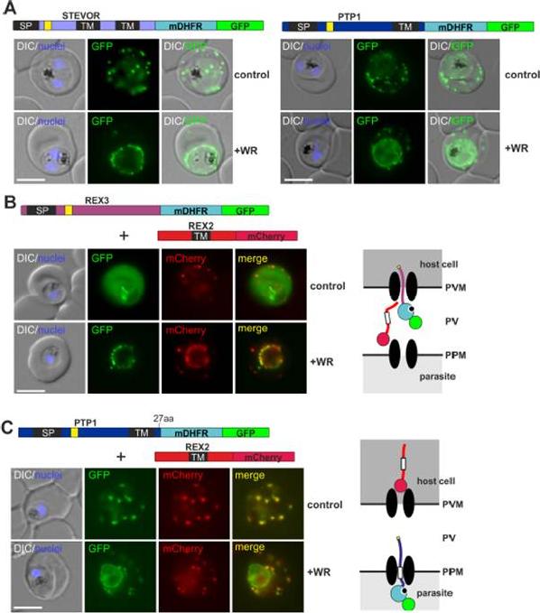
PEXEL TM proteins require translocation for export and some PEXEL proteins can also jam the translocon. (A-C) Representative images of live P. falciparum parasites expressing the constructs shown schematically above each panel (numbers refer to the length of the amino acids sequence between TM and blocking domain). DIC, differential interference contrast. Size bars: 5 μm. A schematic of the co-block (B) and failure to coblock (C) are shown to the right.Mesén-Ramírez P, Reinsch F, Blancke Soares A, Bergmann B, Ullrich AK, Tenzer S, Spielmann T. Stable Translocation Intermediates Jam Global Protein Export in Plasmodium falciparum Parasites and Link the PTEX Component EXP2 with Translocation Activity. PLoS Pathog. 2016 May 11;12(5):e1005618
See original on MMP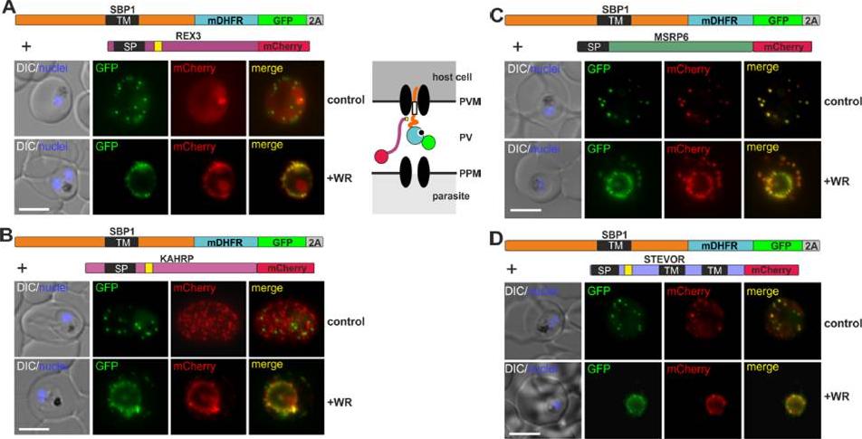
Different types of proteins pass through the same translocon. (A-D) Representative images of live P. falciparum parasites grown in the presence (+WR) or absence of WR (control), expressing the constructs shown schematically above each panel. The skip peptide is indicated by a grey box labelled 2A. The two proteins expressed from the same open reading frame are shown skipped. DIC, differential interference contrast. Size bars: 5 μm. A schematic of the co-block is shown for A. The yellow box indicates the mature PEXEL.Mesén-Ramírez P, Reinsch F, Blancke Soares A, Bergmann B, Ullrich AK, Tenzer S, Spielmann T. Stable Translocation Intermediates Jam Global Protein Export in Plasmodium falciparum Parasites and Link the PTEX Component EXP2 with Translocation Activity. PLoS Pathog. 2016 May 11;12(5):e1005618.
See original on MMP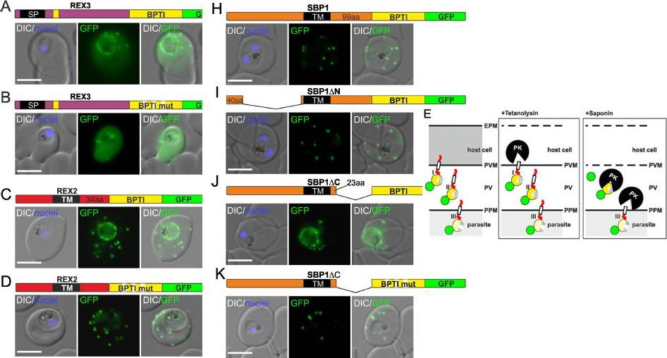
Fusion with the redox sensitive BPTI reveals a second translocation step for TM proteins. (A-D and F-L) Representative images of live P. falciparum parasites expressing the constructs shown schematically above each panel. Hydrophobic regions (SP, signal peptide; TM, transmembrane domain) are in black, the PEXEL motif in yellow. Numbers refer to amino acids (aa). Red boxes labelled C, additional REX2 C-termini. Interrupted yellow box, mutated BPTI (BPTImut). DIC, differential interference contrast. Size bars: 5 μm. (E) Schematic for the protease K (PK) protection assay. Left, intact infected RBC with 3 possibilities (I, II, III) for the location of the fusion construct: I, protein is integral to PVM; II, protein is freely accessible in the PV; III, protein is integral to PPM. Middle, after permeabilisation of the erythrocyte plasma membrane (EPM) with tetanolysin the N-terminus of the construct will be digested if it is in the PVM (I), but remains intact in situation II and III. Right, after permeabilisation of the PVM with saponin, the constructs will be digested if it is in the PVM (I) or the PV (II) but if in the PPM (III), an N-terminally truncated fragment will be generated. Red, exported protein; white box, TM; yellow, BPTI with double cysteine bonds; green, GFP. Mesén-Ramírez P, Reinsch F, Blancke Soares A, Bergmann B, Ullrich AK, Tenzer S, Spielmann T. Stable Translocation Intermediates Jam Global Protein Export in Plasmodium falciparum Parasites and Link the PTEX Component EXP2 withTranslocation Activity. PLoS Pathog. 2016 May 11;12(5):e1005618.
See original on MMPMore information
| PlasmoDB | PF3D7_0936300 |
| GeneDB | PF3D7_0936300 |
| Malaria Metabolic Pathways | Localisation images Pathways mapped to |
| Previous ID(s) | PFI1755c |
| Orthologs | |
| Google Scholar | Search for all mentions of this gene |