PF3D7_0935400 gametocyte development protein 1 (GDV1)
Disruptability [+]
| Species | Disruptability | Reference | Submitter |
|---|---|---|---|
| P. falciparum 3D7 |
Possible |
15760655 | Theo Sanderson, Wellcome Trust Sanger Institute |
| P. falciparum 3D7 |
Possible |
USF piggyBac screen (Insert. mut.) | USF PiggyBac Screen |
Mutant phenotypes [+]
| Species | Stage | Phenotype | Reference | Submitter |
|---|---|---|---|---|
| P. falciparum 3D7 | Asexual |
Transcriptional effect |
http://science.sciencemag.org/content/sci/359/6381/1259.full.pdf
(Knock down)
HP1 levels markedly changed at some genes upon withdrawal of Shield, and transcriptional effects. |
Theo Sanderson, Wellcome Trust Sanger Institute |
| P. falciparum 3D7 | Gametocyte |
Attenuated |
15760655 | Theo Sanderson, Wellcome Trust Sanger Institute |
| P. falciparum 3D7 | Gametocyte |
Attenuated |
http://science.sciencemag.org/content/sci/359/6381/1259.full.pdf
(Knock down)
In the absence of Shield-1, sexual conversion rates decreased markedly. |
Theo Sanderson, Wellcome Trust Sanger Institute |
Imaging data (from Malaria Metabolic Pathways)
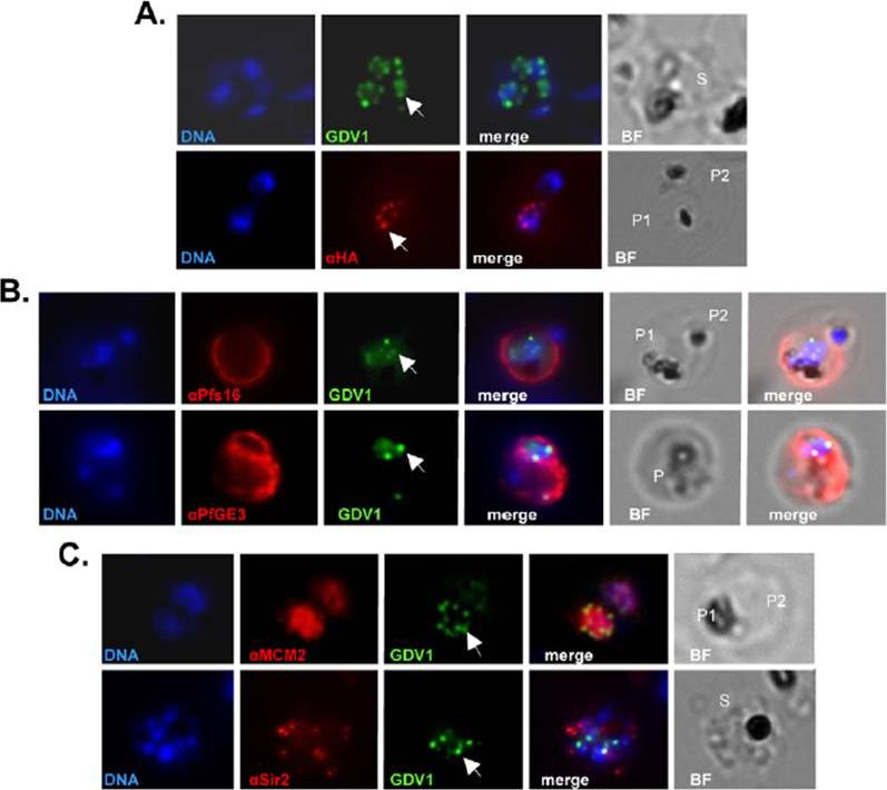
Subcellular localization of PfGDV1. Parasites transformed with GFP- or HA-tagged PfGDV1 were stained with DAPI (DNA stain) and the indicated anti-sera, and then examined by fluorescence microscopy. Images are shown of the DAPI stain (DNA), GFP-tagged PfGDV1 epifluorescence (GDV1), and antibodies specific for HA (aHA), Pfs16 (aPfs16), PfGE3 (aPfGE3), PfMCM2 (aMCM2), and PfSir2 (aSir2). The corresponding merged and bright field (BF) images are included on the right. PfGDV1 expression is indicated with an arrow; locations of parasites in the BF image are indicated with a P for parasite or S for schizont. A) A schizont (S) (Upper) expressing GDV1 and a doubly infected erythrocyte (Lower) with one parasite (P1) expressing HA-tagged PfGDV1 (aHA) and another negative (P2) for anti-HA antibodies. B) Costaining of parasites expressing GDV1 with early gameto-cytogenesis markers. A doubly infected erythrocyte (Upper) with one parasite (P1) positive for GDV1 and aPfs16 and the other (P2) negative for both. An erythrocyte (Lower) infected with a parasite (P) positive for GDV1 and aPfGE3. C) Colocalization of PfGDV1 with nuclear proteins. A doubly infected erythrocyte (Upper) with one parasite in the plane of the image (P1) and the other below (P2). Both P1 and P2 are positive for GDV1 and aMCM2. A schizont (S) (Lower) expressing GDV1 stained with aSir2.Eksi S, Morahan BJ, Haile Y, Furuya T, Jiang H, Ali O, Xu H, Kiattibutr K, Suri A, Czesny B, Adeyemo A, Myers TG, Sattabongkot J, Su XZ, Williamson KC. Plasmodium falciparum gametocyte development 1 (Pfgdv1) and gametocytogenesis early gene identification and commitment to sexual development. PLoS Pathog. 2012;8(10):e1002964.
See original on MMPMore information
| PlasmoDB | PF3D7_0935400 |
| GeneDB | PF3D7_0935400 |
| Malaria Metabolic Pathways | Localisation images Pathways mapped to |
| Previous ID(s) | PFI1710w |
| Orthologs | PKNH_0734100 , PVP01_0734100 , PVX_086950 |
| Google Scholar | Search for all mentions of this gene |