PF3D7_0912400 alkaline phosphatase, putative
Disruptability [+]
| Species | Disruptability | Reference | Submitter |
|---|---|---|---|
| P. falciparum 3D7 |
Possible |
USF piggyBac screen (Insert. mut.) | USF PiggyBac Screen |
Mutant phenotypes [+]
None reported yet. Please press the '+' button above to add one.Imaging data (from Malaria Metabolic Pathways)
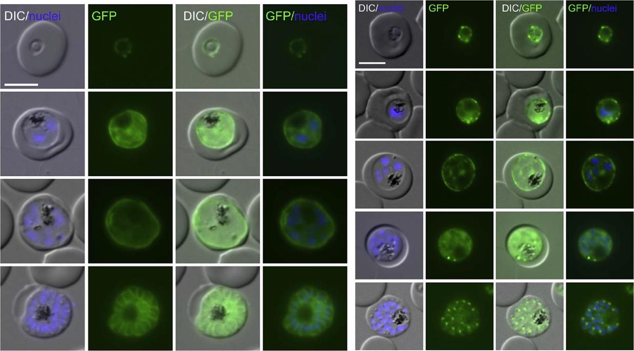
Proteins with TM that are located in the PV. Live cell microscopy images of the endogenously GFP-tagged alkaline phosphatase and EXP3. Left panel: a young and an old schizont are shown to illustrate the change from a staining surrounding the bulk of the still forming merozoites (third row of images) to a ‘bunch of grape’ type pattern where each merozoite is surrounded by GFP signal after the PPM is invaginated (bottom row of images). Right panel: a late trophozoite (third row of images), a young schizont (fourth row of images) and a late schizont (bottom row of images) are shown to demonstrate the change from a peripheral to a parasite internal staining .Khosh-Naucke M, Becker J, Mesén-Ramírez P, Kiani P, Birnbaum J, Fröhlke U, Jonscher E, Schlüter H, Spielmann T. Identification of novel parasitophorous vacuole proteins in P. falciparum parasites using BioID. Int J Med Microbiol. 2017 Jul 27. [Epub ahead of print]
See original on MMP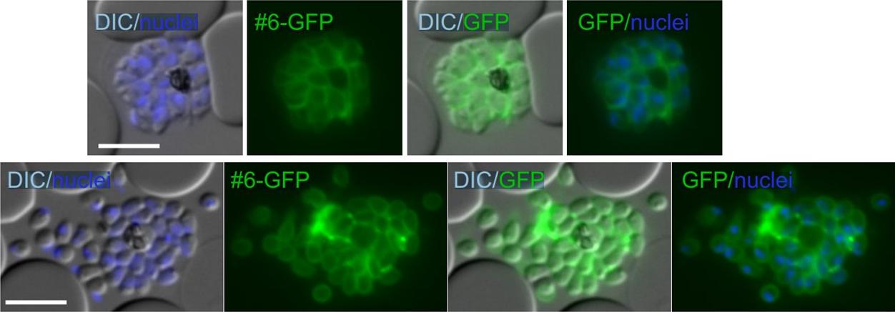
Merozoite surface location of alkaline phosphatase. Live microscopy images of a late schizont showing individual merozoites (top row) or in a ruptured infected red blood cell after release of merozoites (bottom row) in parasites expressing alkaline phosphatase. DIC, differential interference contrast; size bars: 5 mm.Khosh-Naucke M, Becker J, Mesén-Ramírez P, Kiani P, Birnbaum J, Fröhlke U, Jonscher E, Schlüter H, Spielmann T. Identification of novel parasitophorous vacuole proteins in P. falciparum parasites using BioID. Int J Med Microbiol. 2017 Jul 27. [Epub ahead of print]
See original on MMPMore information
| PlasmoDB | PF3D7_0912400 |
| GeneDB | PF3D7_0912400 |
| Malaria Metabolic Pathways | Localisation images Pathways mapped to |
| Previous ID(s) | PFI0605c |
| Orthologs | PBANKA_0813400 , PCHAS_0813700 , PKNH_0710300 , PVP01_0710800 , PVX_099055 , PY17X_0816700 |
| Google Scholar | Search for all mentions of this gene |