PF3D7_0833000 rifin (RIF)
Disruptability [+]
| Species | Disruptability | Reference | Submitter |
|---|---|---|---|
| P. falciparum 3D7 |
Possible |
USF piggyBac screen (Insert. mut.) | USF PiggyBac Screen |
Mutant phenotypes [+]
None reported yet. Please press the '+' button above to add one.Imaging data (from Malaria Metabolic Pathways)
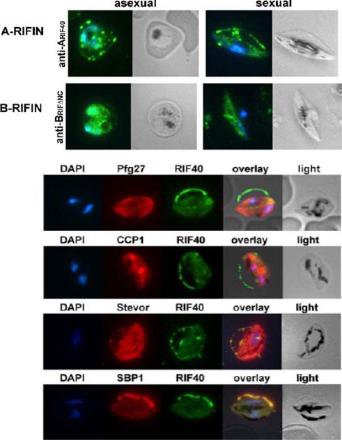
Smears of trophozoite and gametocyte IE were analyzed for RIFIN localization by staining with rat anti-ARIF40 (Rif 40 MAL7P1.217) and mouse anti-BRIFDNC antisera (green) (PFI0050c). Nuclei were stained with DAPI (blue). A type RIFINs were visualized with anti-ARIF40, while B-type RIFINs were detected with anti-BRIFDNC. A-type RIFINs were exported to the host cell in asexual parasites and associated with the Maurer’s clefts (MC), while B-type RIFINs were located mainly intra-parasitically. Immunofluorescence colocalization in stage III gametocytes. Methanol fixed smears of stage III gametocytes were incubated with antibodies against RIFIN and marker proteins. A: rat anti-ARIF40 antibodies (green) and rabbit anti-Pfg27 (cytosol; PF14_0067), mouse anti-CCP1 (PF14_0723), mouse anti-STEVOR or mouse anti-SBP1 (red) (PFE0065w). A-type RIFIN related fluorescence was largely concentrated in a ‘‘string of pearls’’ pattern at the erythrocyte membrane. B-type RIFIN specific staining with anti-BRIFDNC overlapped largely with Pfg27Petter M, Bonow I, Klinkert MQ. Diverse expression patterns of subgroups of the rif multigene family during Plasmodium falciparum gametocytogenesis. PLoS ONE. 2008;3(11):e3779.
See original on MMP
b Co-localization of α-RIF44, STEVOR α-PFL2610w and α-PfMC-2TM-SC (green) with human spectrin (red). c Co-localization of α-RIF44, STEVOR α-PFC0025c and α-PfMC-2TM-CT (green) with SBP1 (red).Bachmann A, Scholz JA, Janßen M, Klinkert MQ, Tannich E, Bruchhaus I, Petter M. A comparative study of the localization and membrane topology of members of the RIFIN, STEVOR and PfMC-2TM protein families in Plasmodium falciparum-infected erythrocytes. Malar J. 2015 Jul 14:274.
See original on MMP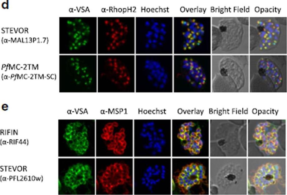
d Co-localization of STEVOR α-MAL13P1.7 or α-PfMC-2TM-SC (green) with the rhoptry marker RhopH2 (red). e Co-localization of α-RIF44 and STEVOR α-PFL2610w (green) with the merozoite surface protein MSP1 (red). Variant surface antigens (VSA).Bachmann A, Scholz JA, Janßen M, Klinkert MQ, Tannich E, Bruchhaus I, Petter M. A comparative study of the localization and membrane topology of members of the RIFIN, STEVOR and PfMC-2TM protein families in Plasmodium falciparum-infected erythrocytes. Malar J. 2015 Jul 14:274.
See original on MMP
Localization of small VSA in infected erythrocytes using confocal immunofluorescence analysis. a Asexual parasites of the 3D7 parasite clone at the trophozoite and schizont stages were fixed with methanol and small VSA localization was visualized using antibodies directed against RIFIN (α-RIF40.2, α-RIF44), STEVOR (α-PFL2610w, α-MAL13P1.7, α-PFC0025c, α-PFA0750w) and PfMC-2TM (α-PfMC-2TM-SC, α-PfMC-2TM-CT) proteins (green). Nuclei were stained with Hoechst33342 (blue).Bachmann A, Scholz JA, Janßen M, Klinkert MQ, Tannich E, Bruchhaus I, Petter M. A comparative study of the localization and membrane topology of members of the RIFIN, STEVOR and PfMC-2TM protein families in Plasmodium falciparum-infected erythrocytes. Malar J. 2015 Jul 14:274.
See original on MMPMore information
| PlasmoDB | PF3D7_0833000 |
| GeneDB | PF3D7_0833000 |
| Malaria Metabolic Pathways | Localisation images Pathways mapped to |
| Previous ID(s) | MAL7P1.217 |
| Orthologs | |
| Google Scholar | Search for all mentions of this gene |