PF3D7_0828800 GPI-anchored micronemal antigen (GAMA)
Disruptability [+]
| Species | Disruptability | Reference | Submitter | |
|---|---|---|---|---|
| P. falciparum 3D7 |
Refractory |
21896773 | Theo Sanderson, Wellcome Trust Sanger Institute | |
| P. falciparum 3D7 |
Refractory |
USF piggyBac screen (Insert. mut.) | USF PiggyBac Screen | |
| P. berghei ANKA |
Possible |
RMgm-484 | Imported from RMgmDB | |
| P. berghei ANKA |
Possible |
RMgm-95 | Imported from RMgmDB | |
| P. berghei ANKA |
Possible |
RMgm-5188 | Imported from RMgmDB | |
Mutant phenotypes [+]
| Species | Stage | Phenotype | Reference | Submitter |
|---|---|---|---|---|
| P. berghei ANKA | Asexual |
No difference |
RMgm-484 | Imported from RMgmDB |
| P. berghei ANKA | Asexual |
No difference |
RMgm-95 | Imported from RMgmDB |
| P. berghei ANKA | Asexual |
No difference |
RMgm-5188 | Imported from RMgmDB |
| P. berghei ANKA | Gametocyte |
No difference |
RMgm-95 | Imported from RMgmDB |
| P. berghei ANKA | Gametocyte |
No difference |
RMgm-5188 | Imported from RMgmDB |
| P. berghei ANKA | Ookinete |
Difference from wild-type |
RMgm-95
Normal production of ookinetes. Possibly a reduced ookinete invasion and traversal of the midgut wall. |
Imported from RMgmDB |
| P. berghei ANKA | Ookinete |
Difference from wild-type |
RMgm-5188
Wild type numbers of ookinetes are produced |
Imported from RMgmDB |
| P. berghei ANKA | Oocyst |
Difference from wild-type |
RMgm-95
Reduced numbers of oocysts: 22% of wild type (9-41%). Oocysts formed are morphologically indistinguishable from wild type oocysts. Sporozoites persist within oocysts at least until day 30 of infection, and fail to produce salivary gland infections. |
Imported from RMgmDB |
| P. berghei ANKA | Oocyst |
Difference from wild-type |
RMgm-5188
(Strongly) reduced oocyst production. Evidence is provided for a defect in the egress of sporozoites from oocysts. |
Imported from RMgmDB |
| P. berghei ANKA | Sporozoite |
Difference from wild-type |
RMgm-95
Sporozoites persist within oocysts at least until day 30 of infection, and fail to produce salivary gland infections, resulting in strongly reduced numbers of salivary gland sporozoites: 0.3% of wild type (0.0-0.7%). No transmission to C57BL/6 mice by bite of infected mosquitoes. |
Imported from RMgmDB |
| P. berghei ANKA | Sporozoite |
Difference from wild-type |
RMgm-5188
(Strongly) reduced oocyst production and strongly reduced numbers of midgut- and salivary gland sporozoites. Evidence is provided for a defect in the egress of sporozoites from oocysts. Sporozoites show strongly reduced motility |
Imported from RMgmDB |
| P. berghei ANKA | Liver |
Difference from wild-type |
RMgm-95
See also the phenotype of sporozoites.No transmission to C57BL/6 mice by bite of infected mosquitoes. Injection of large numbers of midgut sporozoites (500.000) into C57BL/6 mice did not result in a blood stage infection. |
Imported from RMgmDB |
| P. berghei ANKA | Liver |
Difference from wild-type |
RMgm-5188
(Midgut) sporozoites are unable to establish an infection in mice (after intravenous injection). |
Imported from RMgmDB |
Imaging data (from Malaria Metabolic Pathways)
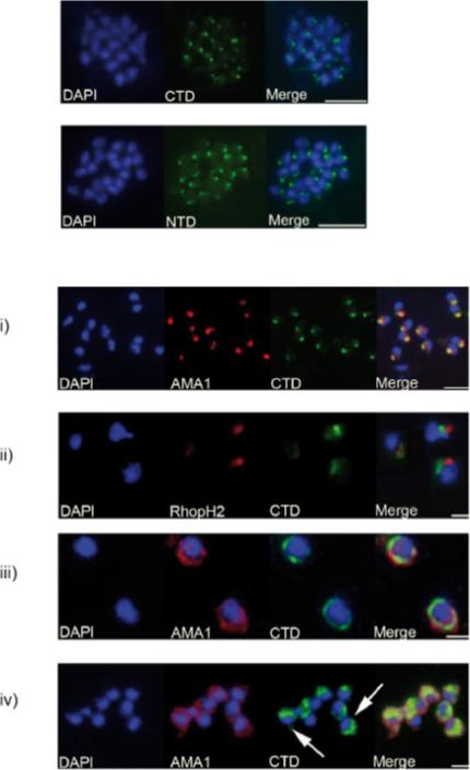
Analyses of the distribution of GAMA in asexual blood stage parasites by immunofluorescence. . Formaldehyde-fixed P. falciparum 3D7 schizonts were probed with mouse anti-CTD (C-terminal domains; top panel) or anti-NTD (N-terminal domains; lower panel) antisera, and an anti-mouse-FITC conjugate (green). Parasite nuclei were stained with DAPI (blue). Staining with both antisera resulted in a strong punctuate pattern, with a single point of fluorescence located away from the nucleus, and consistent with a location in the apical organelles. Scale bars represent 2 mm. (B) Formaldehyde-fixed free P. falciparum merozoites were probed with rabbit anti-CTD antiserum, and either mouse polyclonal anti-AMA1 (PF11_0344)-antibodies (i, iii and iv) or mouse mAb 61.3 (ii). Mouse antibodies were detected with an anti-mouse Alexa Fluor 594 conjugate (red), and rabbit antibodies with an anti-rabbit Alexa Fluor 488 conjugate (green). Parasite nuclei were stained with DAPI (blue). Scale bar in (i) 5 μm, in remainder 2 μm. In (i) and (ii) there is a clear apical localization of AMA1, RhopH2 (PFI1445w) and GAMA, with AMA1 and GAMA appearing to be co-localized; (iii) shows a circumferential localisation of both GAMA and AMA1 on the surface of free merozoites; (iv) shows a ‘cap’-like distribution of GAMA on the surface of free merozoites (arrowed), in contrast to the circumferential distribution of AMA1. Hinds L, Green JL, Knuepfer E, Grainger M, Holder AA. A novel putative GPI-anchored micronemal antigen of Plasmodium falciparum that binds to erythrocytes. Eukaryot Cell. 2009 8:1869-1879. Copyright 2010.
See original on MMP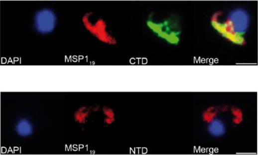
Newly invaded ring-stage parasites were probed with the mouse monoclonal anti-MSP119 (PFI1475w) antibody 1E1, and either rabbit anti-CTD (C-terminal domains; top panel) or anti-NTD (N-terminal domains; lower panel) antisera. Mouse antibodies were detected with an anti-mouse Alexa Fluor 594 conjugate (red) and rabbit antibodies with an anti-rabbit Alexa Fluor 488 conjugate (green). Parasite nuclei were stained with DAPI (blue). Scale bars are 2 μm. There is a clear reactivity of anti-CTD antibodies, but not anti-NTD antibodies, with ring-stage parasites. Anti-CTD and anti-MSP119 signals appear to co-localize (i), suggesting that ‘stub’ part of GAMA that is produced upon shedding of the p42-p37 complex is carried into the host cell and remains associated with the ring-stage parasite. Scale bar 2 μm. Hinds L, Green JL, Knuepfer E, Grainger M, Holder AA. A novel putative GPI-anchored micronemal antigen of Plasmodium falciparum that binds to erythrocytes. Eukaryot Cell. 2009 8:1869-1879. Copyright 2010.
See original on MMP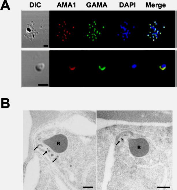
Localization of GAMA in asexual blood stage parasites. (A) GAMA localization using immuno-fluorescence assay. Acetone-fixed P. falciparum 3D7 mature schizonts (top panel) and free merozoites (bottom panel) were probed with rabbit anti-FL (green) and mouse anti-PfAMA1 (microneme marker)(red). Parasite nuclei were stained with DAPI (blue). Scale bars represent 2 μm. (B) GAMA localization using immunoelectron microscopy. The two sections of merozoites in schizont-infected erythrocytes were probed with purified rabbit anti-Tr3 antibody and subsequently by secondary antibody conjugated with gold particles. The arrows indicate the micronemal localization of signals from gold particles. Bars represent 200 nm. Arrows mark micronemes.Arumugam TU, Takeo S, Yamasaki T, Thonkukiatkul A, Miura K, Otsuki H, Zhou H, Long CA, Sattabongkot J, Thompson J, Wilson DW, Beeson JG, Healer J, Crabb BS, Cowman AF, Torii M, Tsuboi T. Discovery of GAMA, a Plasmodium falciparum merozoite micronemal protein, as a novel blood-stage vaccine candidate antigen. Infect Immun. 2011 79(11):4523-32
See original on MMPMore information
| PlasmoDB | PF3D7_0828800 |
| GeneDB | PF3D7_0828800 |
| Malaria Metabolic Pathways | Localisation images Pathways mapped to |
| Previous ID(s) | PF08_0008 |
| Orthologs | PBANKA_0701900 , PCHAS_0936100 , PKNH_1322900 , PVP01_0505600 , PVX_088910 , PY17X_0702200 |
| Google Scholar | Search for all mentions of this gene |