PF3D7_0826700 receptor for activated c kinase (RACK)
Disruptability [+]
| Species | Disruptability | Reference | Submitter |
|---|---|---|---|
| P. falciparum 3D7 |
Refractory |
USF piggyBac screen (Insert. mut.) | USF PiggyBac Screen |
Mutant phenotypes [+]
| Species | Stage | Phenotype | Reference | Submitter |
|---|---|---|---|---|
| P. falciparum 3D7 | Asexual |
Cell cycle arrest |
27732881 (Knock down)
\"Following destabilization, the parasites demonstrate a nearly complete growth arrest at the trophozoite stage. The essential nature of PfRACK1 suggests that the protein itself or the pathways regulated by the protein are potential targets for novel anti-malarial therapeutics.\" |
Theo Sanderson, Francis Crick Institute |
Imaging data (from Malaria Metabolic Pathways)

Immunolocalization of PfRACK in P. falciparum intraerythrocytic stages. (R) ring, (T) trophozoite, and (S) schizont. Methanol-fixed infected erythrocytes were blocked with BSA and incubated with affinity-purified anti-PfRACK antibody. Fluorescence measurements obtained with confocal microscopy correspond to secondary FITC anti-rabbit IgG antibody emission. (A) Phase contrast; (B) immunofluorescence; and (C) merged image.Madeira L, DeMarco R, Gazarini ML, Verjovski-Almeida S, Garcia CR. Human malaria parasites display a receptor for activated C kinase ortholog. Biochem Biophys Res Commun. 2003 306:995-1001. Copyright Elsevier 2009.
See original on MMP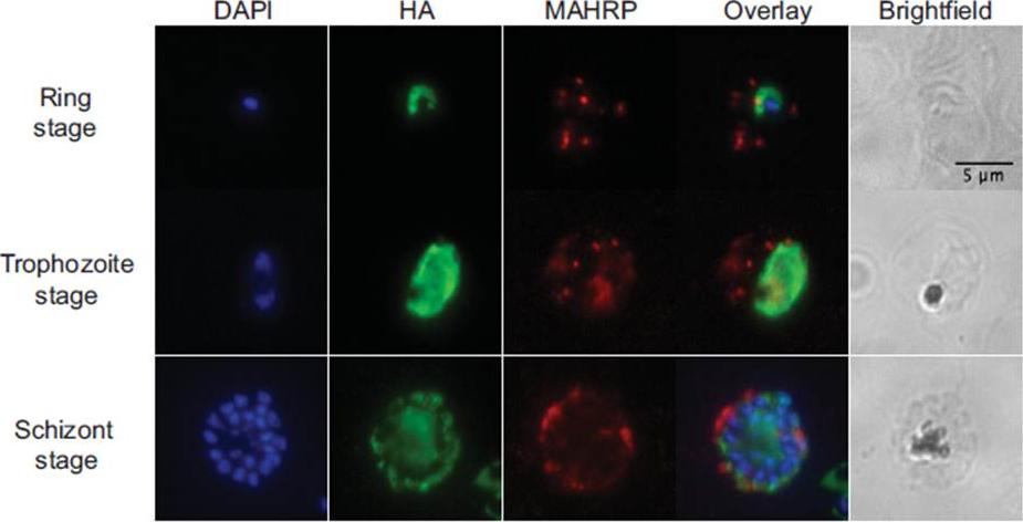
Immunofluorescence. Air-dried thin films of ring (12 hpi), trophozoite (28 hpi) and schizont (44 hpi) stage parasites were fixed in 4% paraformaldehyde, permeabilized with 0.1% Triton X-100, blocked with 3% BSA in phosphate-buffered saline for 1h at RT, incubated with rat α-HA antibody (clone 3F10, 1:50, Roche) at 4oC overnight, and detected with Alexa488-conjugated goat α-rat antibody (1:1000, Invitrogen) for 1h at RT. For PfMAHRP1 co-staining, rabbit α-PfMAHRP1 was added at 1:500 and detected with Alexa555-conjugated goat -rabbit (1:1000, Invitrogen). Slides were mounted with Vectashield anti-fading media containing 4',6-diamidino-2-phenylindole (Vector Laboratories) for nuclear counterstaining. Images were acquired with 100x objectives using a Nikon E800 Microscope. The images were analyzed using ImageJ. PfRACK1 localizes diffusely within the parasite cytoplasm, but not overlapping with the nuclei. we do not observe any exported PfRACK1 in the host cell cytoplasm or in Maurer’s clefts (identified by staining with antibodies directed against PfMAHRP.Blomqvist K, DiPetrillo C, Streva VA, Pine S, Dvorin JD. Receptor for Activated C-Kinase 1 (PfRACK1) is required for Plasmodium falciparum intra-erythrocytic proliferation. Mol Biochem Parasitol. 2016 Oct 9. pii: S0166-6851(16)30129-3.
See original on MMP
fixed schizont stage parasites were stained with rat α-HA antibody and co-stained with rabbit anti-PfERD2 (1:200), rabbit anti-PfBiP (1:500), mouse anti-PfRON4 (1:100), or rabbit anti-PfGAP45 (1:1000). Primary antibodies were detected with Alexa488-conjugated goat α-rat antibody (1:1000) and Alexa555-conjugated goat α-rabbit or goat anti-mouse (1:1000).Blomqvist K, DiPetrillo C, Streva VA, Pine S, Dvorin JD. Receptor for Activated C-Kinase 1 (PfRACK1) is required for Plasmodium falciparum intra-erythrocytic proliferation. Mol Biochem Parasitol. 2016 Oct 9. pii: S0166-6851(16)30129-3.
See original on MMPMore information
| PlasmoDB | PF3D7_0826700 |
| GeneDB | PF3D7_0826700 |
| Malaria Metabolic Pathways | Localisation images Pathways mapped to |
| Previous ID(s) | PF08_0019 |
| Orthologs | PBANKA_0703900 , PCHAS_0934100 , PKNH_1320900 , PVP01_0507600 , PVX_089025 , PY17X_0704200 |
| Google Scholar | Search for all mentions of this gene |