PF3D7_0820700 2-oxoglutarate dehydrogenase E1 component (KDH)
Disruptability [+]
| Species | Disruptability | Reference | Submitter | |
|---|---|---|---|---|
| P. falciparum 3D7 |
Possible |
25843709 | Theo Sanderson, Wellcome Trust Sanger Institute | |
| P. falciparum 3D7 |
Possible |
USF piggyBac screen (Insert. mut.) | USF PiggyBac Screen | |
| P. berghei ANKA |
Possible |
PlasmoGEM (Barseq) | PlasmoGEM | |
Mutant phenotypes [+]
| Species | Stage | Phenotype | Reference | Submitter |
|---|---|---|---|---|
| P. falciparum 3D7 | Asexual |
No difference |
25843709 | Theo Sanderson, Wellcome Trust Sanger Institute |
Imaging data (from Malaria Metabolic Pathways)
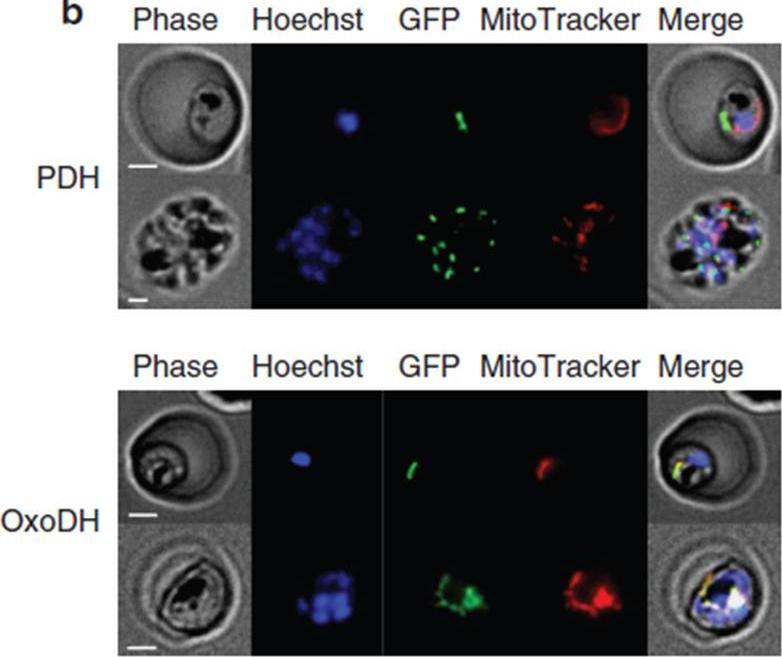
Localization of PfPDH and PfOxoDH within erythrocytes infected with 3D7 parasites. Localization of PfPDH (top panel) and PfOxoDH (bottom panel) in transgenic parasites. The left column shows phase contrast images, followed by fluorescence images of a nuclear dye (Hoechst; blue), GFP-tagged leader sequences of the target enzymes (green), a mitochondrial dye MitoTracker; red) and all four images merged together (right column). Scale bars, 2 mm. The E1-a subunit of PfPDH localizes to a discrete organelle adjacent to the mitochondrion and distinct from the nucleus, consistent with an apicoplast localization (top panel). The truncated E1 subunit of PfOxoDH, which has been predicted to have a mitochondrial localization,colocalizes with MitoTracker when fused to GFP (bottom panel), consistent with a mitochondrial localization.Chan XW, Wrenger C, Stahl K, Bergmann B, Winterberg M, Müller IB, Saliba KJ. Chemical and genetic validation of thiamine utilization as an antimalarial drug target. Nat Commun. 2013 Jun 27;4:2060.
See original on MMP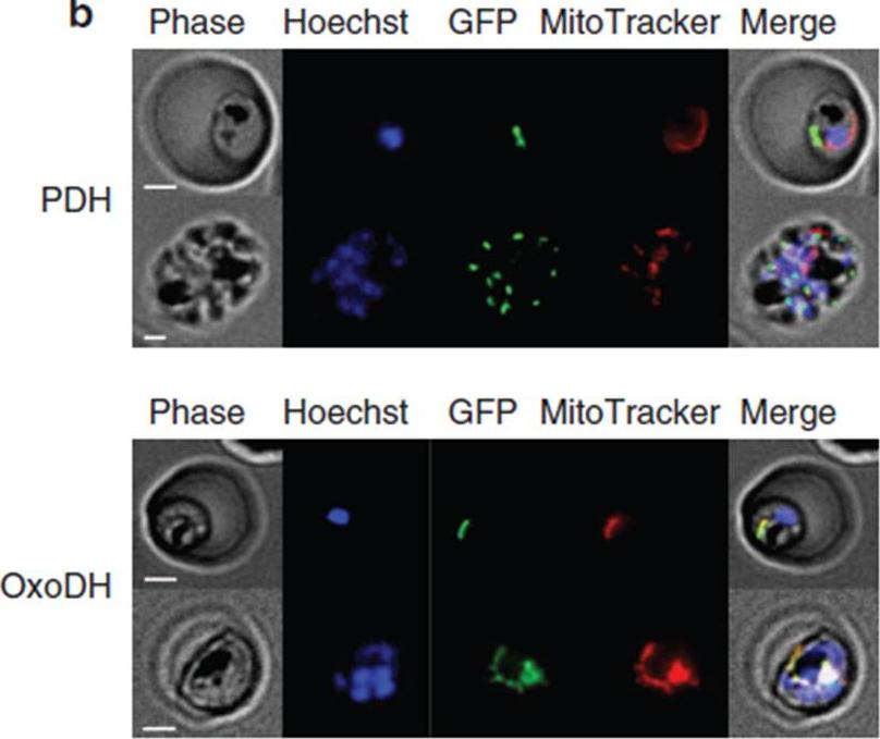
Localization of PfPDH and PfOxoDH within erythrocytes infected with 3D7 parasites. Localization of PfPDH (top panel) and PfOxoDH (bottom panel) in transgenic parasites. The left column shows phase contrast images, followed by fluorescence images of a nuclear dye (Hoechst; blue), GFP-tagged leader sequences of the target enzymes (green), a mitochondrial dye MitoTracker; red) and all four images merged together (right column). Scale bars, 2 mm. The E1-a subunit of PfPDH localizes to a discrete organelle adjacent to the mitochondrion and distinct from the nucleus, consistent with an apicoplast localization (top panel). The truncated E1 subunit of PfOxoDH, which has been predicted to have a mitochondrial localization,colocalizes with MitoTracker when fused to GFP (bottom panel), consistent with a mitochondrial localization.Chan XW, Wrenger C, Stahl K, Bergmann B, Winterberg M, Müller IB, Saliba KJ. Chemical and genetic validation of thiamine utilization as an antimalarial drug target. Nat Commun. 2013 Jun 27;4:2060.
See original on MMPMore information
| PlasmoDB | PF3D7_0820700 |
| GeneDB | PF3D7_0820700 |
| Malaria Metabolic Pathways | Localisation images Pathways mapped to |
| Previous ID(s) | PF08_0045 |
| Orthologs | PBANKA_0710100 , PCHAS_0719200 , PKNH_1314700 , PVP01_0513600 , PVX_089325 , PY17X_0710300 |
| Google Scholar | Search for all mentions of this gene |