PF3D7_0800200 erythrocyte membrane protein 1, PfEMP1 (VAR)
Disruptability [+]
| Species | Disruptability | Reference | Submitter |
|---|---|---|---|
| P. falciparum 3D7 |
Possible |
USF piggyBac screen (Insert. mut.) | USF PiggyBac Screen |
Mutant phenotypes [+]
None reported yet. Please press the '+' button above to add one.Imaging data (from Malaria Metabolic Pathways)
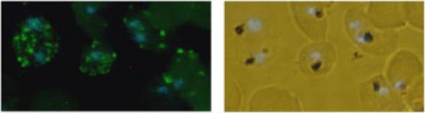
Immunofluorescence analysis of VARC in mature trophozoites. Methanol-fixed parasitized erythrocytes reacted with mouse anti-VARC antibody (1:1000), fluorescein isothiocyanate-conjugated anti-mouse antibodies (1:2000), and DAPI (0.1 mM). The labeled parasites were photographed using a fluorescence microscope (Nikon) at x40 magnification. The left panel shows fluorescence forVARC (green), counterstained for nuclei with DAPI (blue); the right panel shows the corresponding bright field + DAPI.Hora R, Bridges DJ, Craig A, Sharma A. Erythrocytic casein kinase II regulates cytoadherence of Plasmodium falciparum-infected red blood cells. J Biol Chem. 2009 284:6260-9.
See original on MMP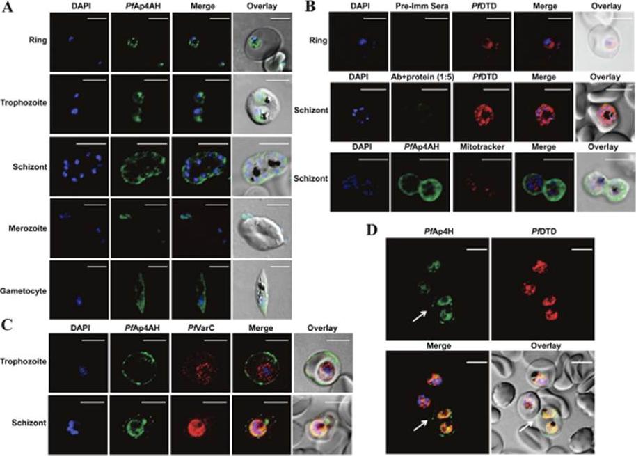
Spatial distribution of P. falciparum Ap4AH during erythrocytic schizogony. Shown are DAPI staining of nucleus in blue and PfAp4AH stained with Alexa 488 in green. (A) Confocal microscopy-data based spatial distribution of PfAp4AH in infected RBCs. PfAp4AH is non-nuclear in blood stages of the parasite and resides in its cytoplasm. (B) Non-mitochondrial localization with various controls is shown. Upper panel shows pre-immune serum (Pre-Imm Sera) control which does not stain the parasite or RBCs. Middle panel shows competitive binding of anti-PfAp4AH antibody to infected cells, where anti-PfAp4AH antibodies were incubated with recombinant PfAp4AH protein in 1:5 ratio. Pf D-tyrosyl-tRNATyr deacylase (DTD) is a cytoplasmic marker. Lower panel shows non-mitochondrial localization where mtochondria are stained in red. (C) RBC membrane localization of PfAp4AH during trophozoite and schizont stages of parasite. VarC is a marker for infected RBC membrane localization. (D) A field view of anti-PfAp4AH antibody staining of infected RBCs. Significant fraction of cells (~50%) showed membrane localization of PfAp4AH - here cell is marked with white arrow. Uninfected RBCs (without DAPI and PfDTD staining here) are unstained. White scale bar in confocal figures is of 5 μmSharma A, Yogavel M, Sharma A. Structural and functional attributes of malaria parasite diadenosine tetraphosphate hydrolase. Sci Rep. 2016 Feb 1;6:19981.
See original on MMPMore information
| PlasmoDB | PF3D7_0800200 |
| GeneDB | PF3D7_0800200 |
| Malaria Metabolic Pathways | Localisation images Pathways mapped to |
| Previous ID(s) | MAL8P1.167, PF08_0141 |
| Orthologs | |
| Google Scholar | Search for all mentions of this gene |