PF3D7_0731500 erythrocyte binding antigen-175 (EBA175)
Disruptability [+]
| Species | Disruptability | Reference | Submitter |
|---|---|---|---|
| P. falciparum 3D7 |
Possible |
12672957 | Theo Sanderson, Wellcome Trust Sanger Institute |
| P. falciparum 3D7 |
Possible |
USF piggyBac screen (Insert. mut.) | USF PiggyBac Screen |
Mutant phenotypes [+]
| Species | Stage | Phenotype | Reference | Submitter |
|---|---|---|---|---|
| P. falciparum 3D7 | Asexual |
Attenuated |
12672957 | Theo Sanderson, Wellcome Trust Sanger Institute |
Imaging data (from Malaria Metabolic Pathways)
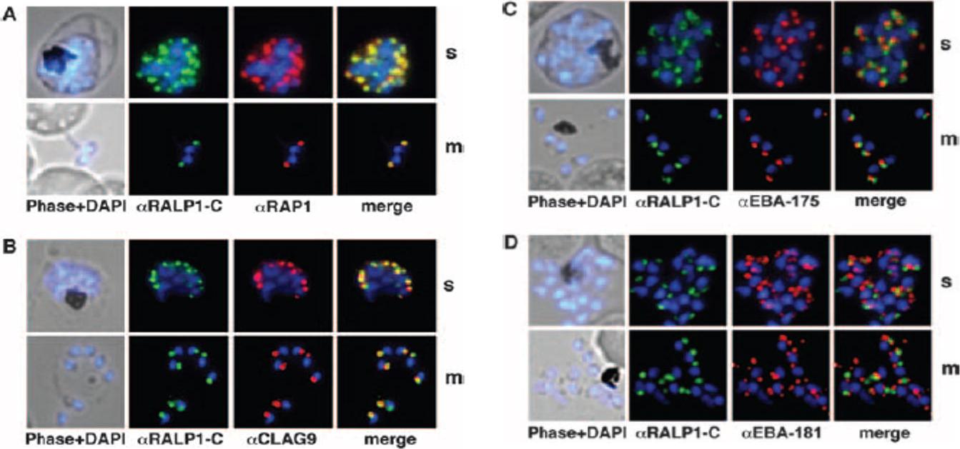
Colocalization studies of RALP1 with rhoptry and microneme marker proteins. (A and B) RALP1-C-specific antibodies (green) colocalize with the rhoptry protein RAP1 (red) (A) and predominantly colocalize with CLAG9 a rhoptry-specific marker, (red) (B) in fixed schizonts (s) and free merozoites (m) using RAP1- and CLAG9-specific antibodies, respectively. (C and D) RALP1-C-specific antibodies (green) visualize a different compartment within the parasite than the microneme marker proteins EBA-175 (red) (C) and EBA-181 (red) (D), as is evident in the merge of the two fluorescence photomicrographs. Nuclei were stained blue (DAPI).Haase S, Cabrera A, Langer C, Treeck M, Struck N, Herrmann S, Jansen PW, Bruchhaus I, Bachmann A, Dias S, Cowman AF, Stunnenberg HG, Spielmann T, Gilberger TW. Characterization of a conserved rhoptry-associated leucine zipper-like protein in the malaria parasite Plasmodium falciparum. Infect Immun. 2008 76:879-87.
See original on MMP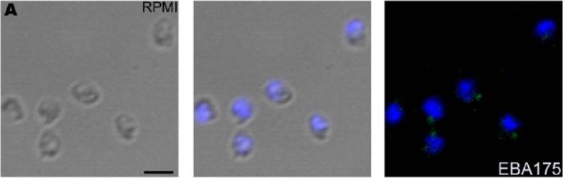
Expression of microneme protein EBA175 on surface of P. falciparum merozoites isolated in RPMI1640 medium (RPMI). detected by IFA using anti-EBA175 rabbit sera followed by FITC-conjugated anti-rabbit IgG goat sera (green). Nuclear DNA was counterstained with DAPI (blue). Bright field, bright field merged with DAPI staining and merged fluorescence images (DAPI and FITC) are shown. Black bar indicates 2 mm.Singh S, Alam MM, Pal-Bhowmick I, Brzostowski JA, Chitnis CE. Distinct external signals trigger sequential release of apical organelles during erythrocyte invasion by malaria parasites. PLoS Pathog. 2010 Feb 5;6(2):e1000746.
See original on MMP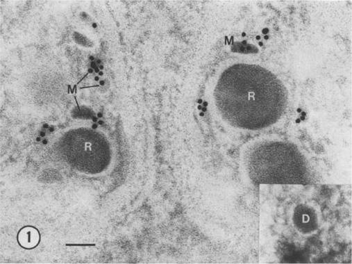
lmmunoelectron micrograph showing the presence of EBA-peptide 4 in micronemes (M) of P. falciparum merozoites. No gold particles are seen associated with rhoptries (R). Insert: dense granule (D) without gold particles. Bar = 100 nm.Sim BK, Toyoshima T, Haynes JD, Aikawa M. Localization of the 175-kilodalton erythrocyte binding antigen in micronemes of Plasmodium falciparum merozoites. Mol Biochem Parasitol. 1992 51:157-9. Copyright Elsevier 2010.
See original on MMP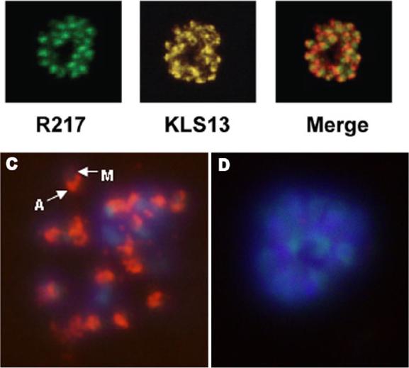
Dual immunofluorescent analyses showing apical staining of mature P. falciparum (FVO strain) schizont with EBA-175 RII specific mAb R217 used at 10 mg/mL and rabbit polyclonal sera KLS13 against baculovirus expressed EBA-175 RII. These mAbs gave a characteristic apical punctate fluorescence pattern in late stage schizonts.C) Schizont stained with mAb R217 (used at 2.34 mg/mL), M: merozoite; A: apical end of the merozoite expressing EBA175, D) schizont stained with anti-PfCSP mAb 2A10 (used at 68 mg/mL). The nuclei were stained with DAPI. Sim BK, Narum DL, Chattopadhyay R, Ahumada A, Haynes JD, Fuhrmann SR, Wingard JN, Liang H, Moch JK, Hoffman SL. Delineation of stage specific expression of Plasmodium falciparum EBA-175 by biologically functional region II monoclonal antibodies. PLoS One. 2011 Apr 14;6(4):e18393.
See original on MMP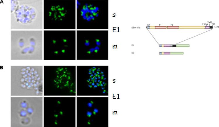
Localization of EBA-175 GFP by fluorescence microscopy in unfixed parasites. A and B, using fluorescence of the GFP reporter protein E1 and E2 distribution (green) is restricted to the apical pole within the parasite. The nucleus is stained with DAPI (blue). EBA-175 is localized in micronemes.Treeck M, Struck NS, Haase S, Langer C, Herrmann S, Healer J, Cowman AF, Gilberger TW. A conserved region in the EBL proteins is implicated in microneme targeting of the malaria parasite Plasmodium falciparum. J Biol Chem. 2006 281:31995-2003.
See original on MMP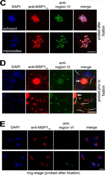
EBA-175 is shed at or around the point of erythrocyte invasion and retains region VI. (C) IFA images of an acetone-fixed segmented 3D7 schizont and free merozoites dual labeled after fixation with mAb 1E1 (anti-MSP119; red) and anti–region VI antibodies (green). The punctate, apical pattern obtained with the latter is typical of microneme staining. (D) EBA-175 is discharged onto the apical surface of free merozoites. Merozoites released from 3D7 schizonts in the presence of anti–region VI antibodies were washed, fixed, and probed with FITC anti-mouse IgG to detect bound anti–region VI antibodies (green), followed by mAb 1E1 (anti-MSP119; red). Many free merozoites exhibit a strong apical FITC signal (thin arrows), whereas residual intact schizonts containing merozoites that were not accessible to the anti–region VI antibodies show only background FITC labeling (thick arrows). No signal was associated with merozoites released in the presence of preimmune mouse antisera (unpublished data). (E) IFA images of newly invaded (≤3-h-old) ring-stage 3D7 parasites probed with anti–region VI antibodies after acetone fixation. Most rings did not react at all with the antibodies. Nuclei were stained with DAPI (blue). Bars, 5 μm.O'Donnell RA, Hackett F, Howell SA, Treeck M, Struck N, Krnajski Z, Withers-Martinez C, Gilberger TW, Blackman MJ. Intramembrane proteolysis mediates shedding of a key adhesin during erythrocyte invasion by the malaria parasite. J Cell Biol. 2006 174:1023-33.
See original on MMP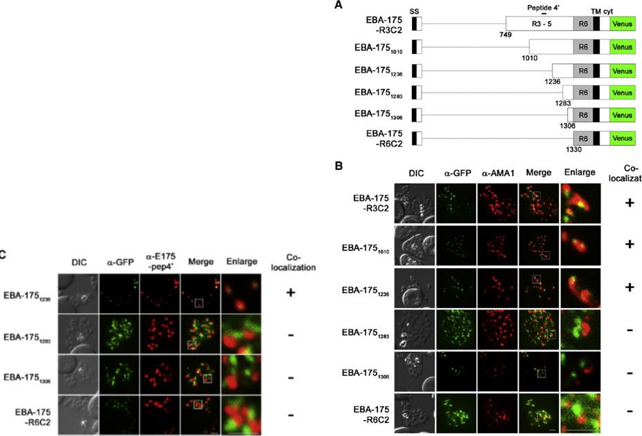
IFA detection of parasites expressing serially truncated recombinant EBA-175. (A) Schematic structures of recombinant EBA-175 proteins; EBA-175-R3C2, EBA-1751010, EBA-1751236, EBA-1751283, EBA-1751306 or EBA-175-R6C2. Signal sequence (SS), regions 1 to 6 (R1, R2, R3–5 and R6), transmembrane region (TM) and cytoplasmic tail (cyt) are indicated. Bar above the scheme indicates the target region of anti-EBA-175-pep4′ antibody (aa 1089–1108). (B and C) Parasites were stained with anti-GFP (α-GFP, green) and anti-AMA1 (panel B; α-AMA1, red) or anti-EBA-175-pep4′ (panel C; α-E175-pep4′, red). Scale bar indicates 2 μm. DIC is a differential interference contrast image. Status of the co-localization with a microneme marker is shown at the right side of each row. (+) or (−) indicate that two signal co-localized or not co-localized.Sakura T, Yahata K, Kaneko O. The upstream sequence segment of the C-terminal cysteine-rich domain is required for microneme trafficking of Plasmodium falciparum erythrocyte binding antigen 175. Parasitol Int. 2012 62(2):157-64
See original on MMP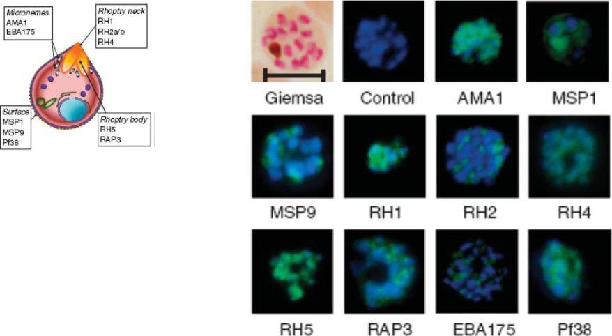
Indirect immunofluorescence images of P. falciparum schizonts stained with rabbit IgG (green) induced by 10 viral-vectored vaccines expressing malaria antigens, and negative control vectors lacking a malaria antigen. Nuclei are counterstained with 4,6-diamidino-2-phenylindole (blue). All sera were tested against 3D7 strain parasites, with the exception of anti-PfRH1 for which FVO strain parasites were used. All images to same scale as Giemsa stained image (top left, on which scale bar indicates 5 μm).Douglas AD, Williams AR, Illingworth JJ, Kamuyu G, Biswas S, Goodman AL, Wyllie DH, Crosnier C, Miura K, Wright GJ, Long CA, Osier FH, Marsh K, Turner AV, Hill AV, Draper SJ. The blood-stage malaria antigen PfRH5 is susceptible to vaccine-inducible cross-strain neutralizing antibody. Nat Commun. 2011 2:601.
See original on MMP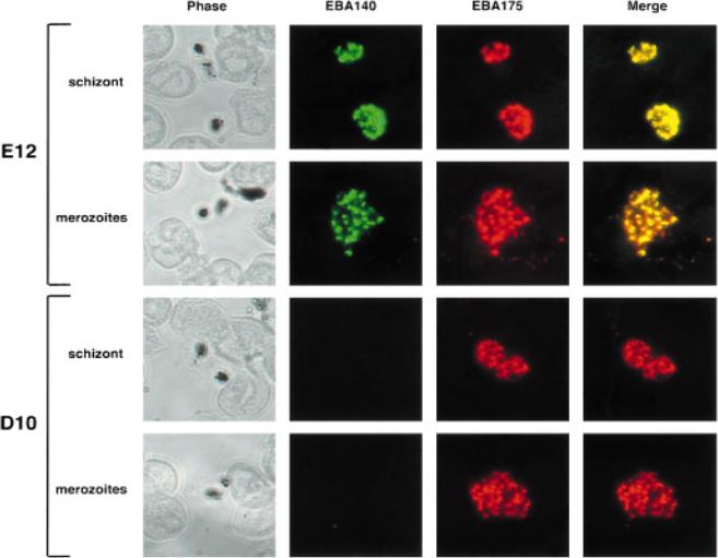
The EBA140 and EBA175 proteins colocalize in in the micronemes,of merozoites. Smears of schizonts or free merozoites of the D10 and E12 parasites are shown. Parasites were reacted with a mixture of anti-EBA140 and anti-EBA175 antibodies, followed by a mixture of FITC labelled anti-mouse and rhodamine-labelled anti-rabbit antibodies. ‘Phase’ shows the brightfield image, whereas ‘merge’ shows the red and green images overlaid.Thompson JK, Triglia T, Reed MB, Cowman AF. A novel ligand from Plasmodium falciparum that binds to a sialic acid-containing receptor on the surface of human erythrocytes. Mol Microbiol. 2001 41:47-58. Copyright John Wiley & Sons Ltd. 2010.
See original on MMP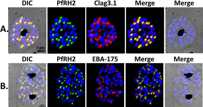
Localization of PfRH2a/b by immunofluorescence confocal microscopy. (A) 3D7 schizonts were dual labeled with anti-rPfRH240 mice sera and anti-clag3.1 rabbit sera. Mature schizonts immunolabeled with anti-rPfRH240 were stained with Alexa 488 linked anti-mouse IgG secondary antibody (green). Schizonts labeled with anti-clag3.1 rabbit sera were stained with Alexa 594 linked anti-rabbit IgG secondary antibody (red). (B) 3D7 mature schizonts were dual labeled with anti-rPfRH240 mouse sera and anti-EBA175 rabbit sera. Schizonts labeled with anti-EBA-175 antibodies were stained with Alexa 594 linked anti-rabbit IgG secondary antibody (red). PfRH2a/b co-localizes with the known rhoptry bulb marker protein, clag3.1 demonstrating that it is localized in the rhoptries, and not with the microneme marker protein, EBA-175.Sahar T, Reddy KS, Bharadwaj M, Pandey AK, Singh S, Chitnis CE, Gaur D. Plasmodium falciparum reticulocyte binding-like homologue protein 2 (PfRH2) is a key adhesive molecule involved in erythrocyte invasion. PLoS One. 2011 6(2):e17102.
See original on MMP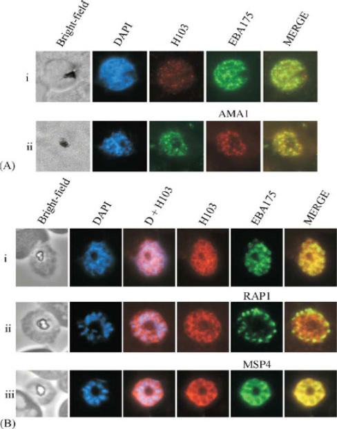
(A) Immunofluorescence of early schizont parasites indicated that H103 partially co-localises with EBA175 (i) and AMA1 (ii) in the micronemes. DAPI staining indicates that the parasites are early schizonts. (B) Immunofluorescence of late stage schizont parasites using antibodies to H103, EBA175 (i), RAP1 (ii) and MSP4 (iii) indicated that at this stage of the asexual cycle H103 was located on the merozoite surface. H103 did not co-localise with the microneme protein EBA175 or the rhoptries protein RAP1 at this stage of the cycle. DAPI staining shows the location of DNA and signifies that the parasites are fully segmented schizonts.Pearce JA, Mills K, Triglia T, Cowman AF, Anders RF. Characterisation of two novel proteins from the asexual stage of Plasmodium falciparum, H101 and H103. Mol Biochem Parasitol. 2005 139:141-51. Copyright Elsevier 2009.
See original on MMP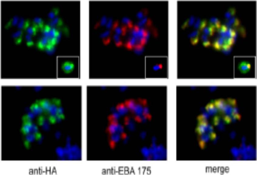
A. IFA of drug-selected 3D7 parasites harbouring the AMA_M3 construct. The transgenic PfAMA1/DIII-HA product detected by anti-HA mAb 3F10 (green) co-localises with the microneme protein EBA-175 (red) in mature segmented schizonts. The inset shows similar staining of a free merozoite, where some translocation of PfAMA1/DIIIHA onto the merozoite surface is detectable as is normal for endogenous PfAMA1. B. IFA of drug-selected 3D7 parasites harbouring the AMA_ins construct. The transgene product detected by anti-HA mAb 3F10 (green) co-localises correctly with the microneme protein EBA-175 (red) in mature segmented schizonts.Olivieri A, Collins CR, Hackett F, Withers-Martinez C, Marshall J, Flynn HR, Skehel JM, Blackman MJ. Juxtamembrane shedding of Plasmodium falciparum AMA1 is sequence independent and essential, and helps evade invasion-inhibitory antibodies. PLoS Pathog. 2011 7(12):e1002448.
See original on MMP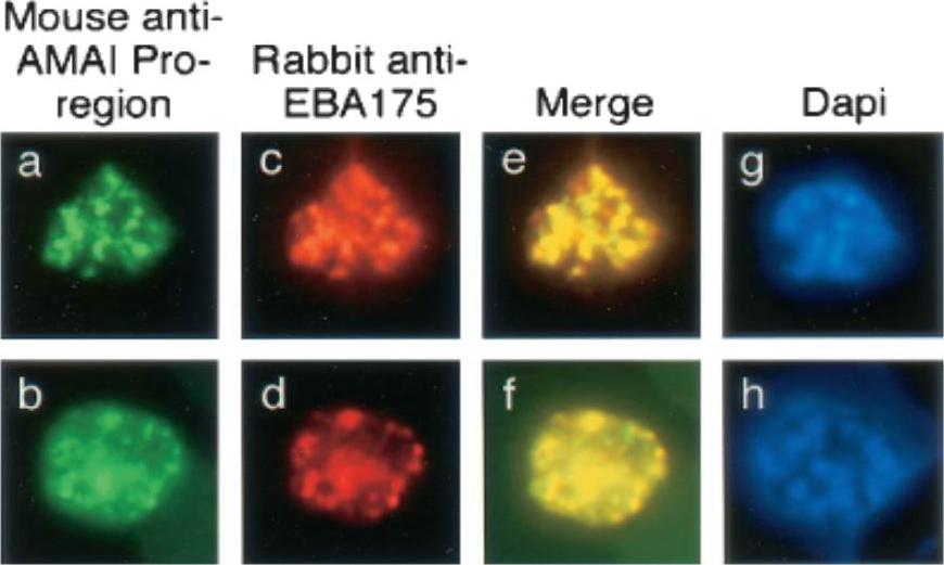
Single and dual indirect immunofluorescence images showing the same subcellular localization of AMA1 and EBA-175 in two mature schizonts of P. falciparum. (a to d) FITC-labeled MAb 5G8 against the PfAMA1 pro-region (a and b) and rhodamine-labeled anti-EBA175 (c and d). (e and f) Merged images show full-length AMA1 colocalized with EBA175. (g and h) DAPI-stained parasites included to show the stage of merozoite development. MAb 5G8, which specifically recognizes the unprocessed form of AMA1, showed a punctate pattern characteristic of the apical organelles of merozoites (a and b). Anti-EBA175 antibodies showed the same pattern (c and d), and merging of the images from the FITC and rhodamine channels suggested that these two proteins were colocalized in the same subcellular compartment, the micronemes.Healer J, Crawford S, Ralph S, McFadden G, Cowman AF. Independent translocation of two micronemal proteins in developing Plasmodium falciparum merozoites. Infect Immun. 2002 70:5751-8.
See original on MMP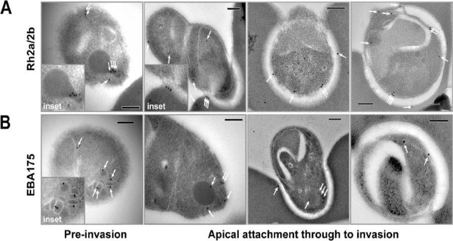
Immuno-electron microscopy localisation of PfRh2a and PfRh2b during merozoite invasion of human erythrocytes. (A) The PfRh2a/2b proteins localise to the rhoptry neck and moving junction of merozoites. Transmission electron micrographs of pre- and mid-invasion merozoites showing PfRh2a/b-specific immunogold labelling (white arrows). At the pre-invasion stage, PfRh2a/b localises to the rhoptry neck (inset), while during mid-invasion, PfRh2a/b localises both to the moving junction and to the merozoite surface within the invasion pit. Scale bar = 0.2 mm. (B) The EBA-175 protein localises to the micronemes and apical tip of merozoites. At the pre-invasion stage, EBA-175 is located within micronemes (inset), while during mid-invasion, EBA-175 can be located either at the apical tip or within the invasion pit. Scale bar = 0.2 mm.Triglia T, Chen L, Lopaticki S, Dekiwadia C, Riglar DT, Hodder AN, Ralph SA, Baum J, Cowman AF. Plasmodium falciparum merozoite invasion is inhibited by antibodies that target the PfRh2a and b binding domains. PLoS Pathog. 2011 (6):e1002075.
See original on MMP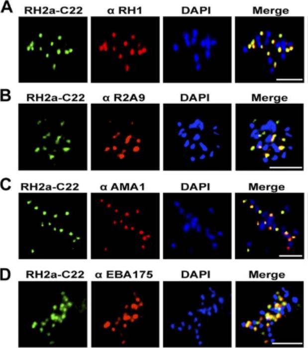
An immunofluorescence assay was performed on late-schizont-stage W2mef parasites stained for RH2a using the anti-RH2a MAb C22 and costained either with an antibody against the rhoptry neck marker RH1 (A), rabbit polyclonal antibody R2A9, recognizing both RH2a and RH2b (B), or an antibody against the micronemal marker AMA1 (C) or EBA175 (D). Bars, 5 mm. PfRH2a showed a punctate pattern at the apical tip of the merozoite in schizonts and appeared to colocalize with PfRH1 in W2mef and 3D7 parasites, strongly suggesting that RH2a is located at the rhoptry neck.Gunalan K, Gao X, Liew KJ, Preiser PR. Differences in erythrocyte receptor specificity of different parts of the Plasmodium falciparum reticulocyte binding protein homologue 2a. Infect Immun. 2011 79:3421-30.
See original on MMP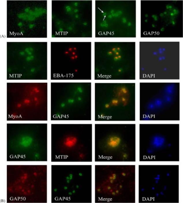
Components of the P. falciparum merozoite glideosome localize to the periphery of the parasite. (A) Indirect-immunofluorescence of individual glideosome components in late blood-stage parasites: Thin smears of P. falciparum infected erythrocytes were probed with anti-glideosome sera. The arrow indicates a gap in peripheral staining at the end of the merozoite, suggestive of an inner membrane complex (IMC) localization. (B) Indirect-immunofluorescence of late blood-stage parasites co-stained with antisera to other glideosome components and PfEBA-175: Glideosome antisera were used in co-immunofluorescence assays to assess co-localization. Anti-PfEBA-175 antisera recognize an invasion ligand that is localized to the micronemes, and is used as a marker for the apical end of the merozoite.Jones ML, Kitson EL, Rayner JC. Plasmodium falciparum erythrocyte invasion: a conserved myosin associated complex. Mol Biochem Parasitol. 2006 147:74-84. Copyright Elsevier 2009.
See original on MMP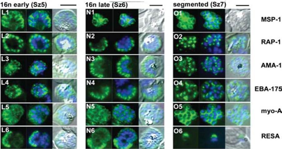
The appearance of Plasmodium falciparum schizonts of the ITO4 line at different times of development. The time of sampling from invasion and the number of nuclei per schizont are indicated. Antibodies to proteins associated with invasion related organelles or the parasite periphery (rhoptries : RAP-1; micronemes: AMA-1, EBA-175; dense granules: RESA; parasite periphery: MSP-1, myo-A) were chosen to follow the development and assembly of merozoites in maturing schizonts.Margos G, Bannister LH, Dluzewski AR, Hopkins J, Williams IT, Mitchell GH. Correlation of structural development and differential expression of invasion-related molecules in schizonts of Plasmodium falciparum. Parasitology. 2004 129:273-87. Copyright Cambridge University Press Journals 2011
See original on MMP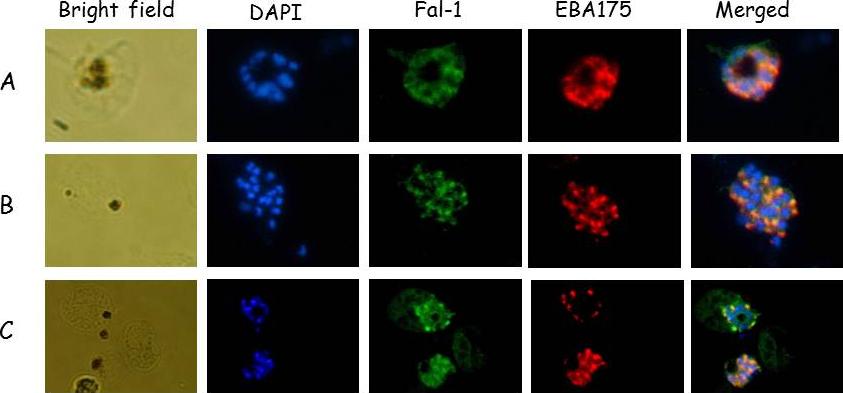
Falcipain 1 and EBA-175 (a merozoite microneme protein) co-localized at the schizont stage. Immunofluorescence images of fixed 3D7 P. falciparum parasites. (A) Merozoites costained with anti-F1.4 and anti-EBA-175 antibodies. (B) Schizonts costained with anti-F1.4 and anti-EBA-175 antibodies. (C) Merozoites costained with anti-F1.4 and anti-MSP-119 antibodies.Kumar A, Kumar K, Korde R, Puri SK, Malhotra P, Singh Chauhan V. Falcipain-1, a Plasmodium falciparum cysteine protease with vaccine potential. Infect Immun. 2007 75:2026-34.
See original on MMP
Localization of PfMA by Immuno-electron microscopy. Ultra thin sections of P. falciparum parasites at schizont/merozoite stages were labelled with mouse anti-PfMA or anti-EBA-175 antibodies followed with anti-mouse secondary antibody conjugated with 15nm colloidal gold particle. (A) Labelling with the PfMA antibody was confined only to the micronemes (arrows) in the apical end of the free merozoite and was absent from the rhoptry (Rh) or nucleus (Nu). (B, C) Similarly PfMA staining in the schizonts was observed only in the micronemes and not in the rhoptries. (D, E) A similar pattern of staining in the schizonts was observed with the micronemal marker EBA-175.Hans N, Singh S, Pandey AK, Reddy KS, Gaur D, Chauhan VS. Identification and Characterization of a Novel Plasmodium falciparum Adhesin Involved in Erythrocyte Invasion. PLoS One. 2013 Sep 13;8(9):e74790.
See original on MMP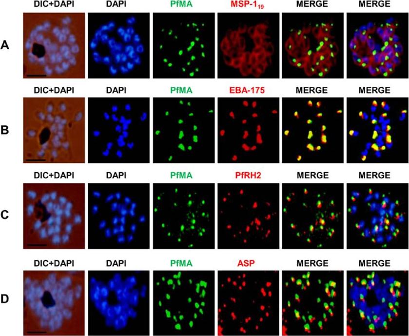
PfMA localization in schizont stages analyzed by fluorescence microscopy. Sub-cellular localization of PfMA was studied by co-immunostaining with surface protein (A), microneme (B), rhoptry (C, D) resident proteins. P. falciparum schizonts were co-immunostained with mouse anti-PfMA (green) and rabbit antibodies against one of the 4 marker proteins (EBA175/PfRH2, ASP, MSP-119) (red). The nuclei of schizont were stained with DAPI (blue) and slides were visualized by fluorescence microscope. All apical marker proteins and PfMA showed punctate staining in schizonts. PfMA was localized in the micronemes as it signal costained with micronemal marker EBA-175. The scale bar indicates 2 μm.Hans N, Singh S, Pandey AK, Reddy KS, Gaur D, Chauhan VS. Identification and Characterization of a Novel Plasmodium falciparum Adhesin Involved in Erythrocyte Invasion. PLoS One. 2013 8(9):e74790.
See original on MMP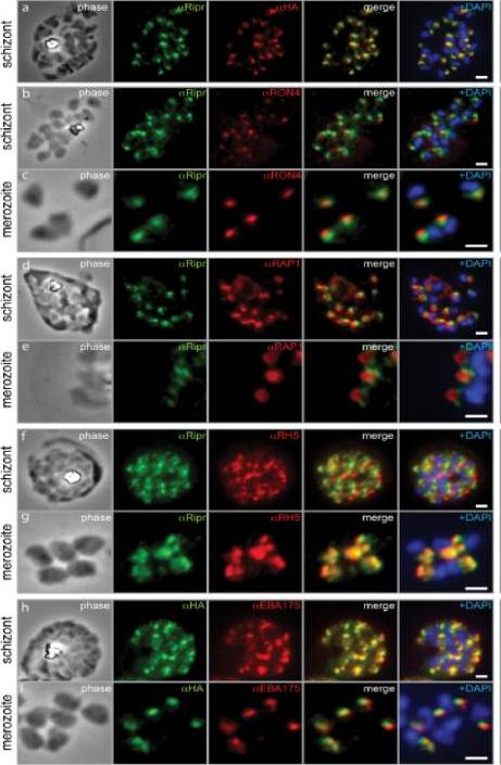
PfRipr localizes to the apical end of merozoites. a) Rabbit polyclonal anti-PfRip antibody (anti-PfRipr/1) recognizes HA-tagged PfRiprHA. b) PfRipr does not co-localize with the rhoptry neck protein RON4 in schizonts. c) PfRipr does not co-localize with the rhoptry neck protein RON4 in merozoites. d) PfRipr does not co-localize with the rhoptry bulb protein, RAP1 in schizonts. e) PfRipr does not co-localize with the rhoptry bulb protein, RAP1 in merozoites. f) PfRipr partially co-localizes with PfRh5 in the schizonts. g) PfRipr mainly co-localizes with PfRh5 in purified merozoites. h) PfRipr co-localizes with the micronemal marker, EBA175, in schizonts. i) PfRipr does not co-localize with the micronemal marker, EBA175, in merozoites.Chen L, Lopaticki S, Riglar DT, Dekiwadia C, Uboldi AD, Tham WH, O'Neill MT, Richard D, Baum J, Ralph SA, Cowman AF. An EGF-like protein forms a complex with PfRh5 and is required for invasion of human erythrocytes by Plasmodium falciparum. PLoS Pathog. 2011 7(9):e1002199.
See original on MMP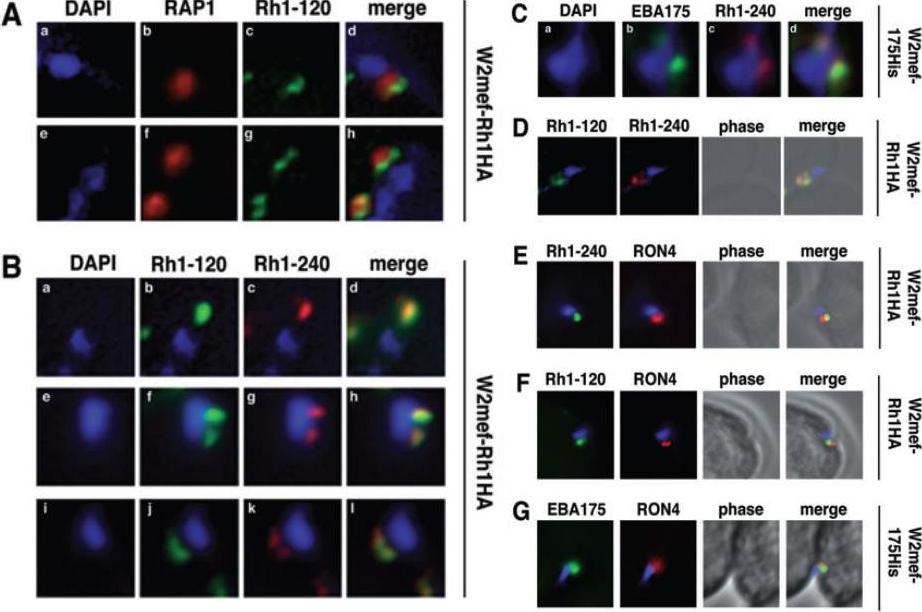
Schizont-processed Rh1-240 colocalizes with Rh1-120 at the apical tip and tight junction of the merozoite. A. The Rh1-120 protein is located at the apical tip and tight junction of merozoites. Free merozoites from the W2mef-Rh1HA line were dual stained with a McAb to RAP1 in the body of the rhoptry and with rat anti-HA antibodies to the tagged 120 kDa Rh1. Anti-HA staining is seen either apical to the RAP1 staining (top panels) or at both sides but posterior to RAP1 staining indicating tight junction staining (bottom panels). Nuclei were stained with DAPI. B. The Rh1-120 protein colocalizes with the Rh1-240 protein at the apical tip and tight junction of merozoites. Free merozoites from the W2mef-Rh1HA line were dual-stained with rat anti-HA antibodies to the tagged 120 kDa Rh1 and with a rabbit antibody (R515) to the 240 kDa Rh1. Colocalization is seen at the apical tip (panels a–d) and at the tight junction (panels e–l) of individual merozoites. W2mef-175His line C. The Rh1-240 protein colocalizes with EBA-175 MAL7P1.176 at the tight junction of merozoites. Free merozoites were dual-stained with mouse anti-His antibodies to tagged EBA-175 and with a rabbit antibody to the 240 kDa Rh1. EBA-175 and Rh1-240 appear to colocalize at the tight junction. D. Free merozoites were dual-stained with rat anti-HA antibodies to the tagged 120 kDa Rh1 and with a rabbit antibody to the 240 kDa Rh1. Simultaneous staining of both the apical tip and the tight junction of a merozoite is seen. E–G. Merozoites were trapped during invasion using cytochalasin D. Rh1-240 was stained with R515, Rh1-120 with rat anti-HA and EBA-175 with mouse anti-His antibodies. PfRON4 was stained either with a mouse antibody (E) or with a rabbit antibody (F and G). Triglia T, Tham WH, Hodder A, Cowman AF. Reticulocyte binding protein homologues are key adhesins during erythrocyte invasion by Plasmodium falciparum. Cell Microbiol. 2009 11:1671-87. Copyright John Wiley & Sons Ltd. 2010.
See original on MMP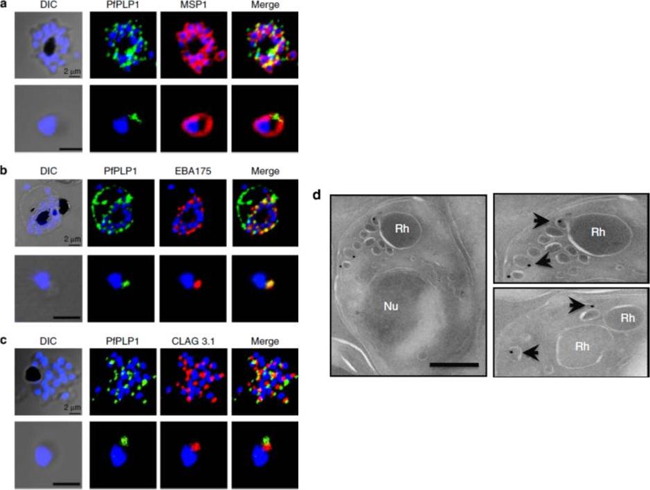
Subcellular localization of PfPLP1 in schizonts and merozoites of P. falciparum. (a) Co-immunostaining of PfPLP1 (green) with merozoite surface protein MSP1 (red). (b) Co-immunostaining of PfPLP1 (green) with micronemal protein EBA-175 (red). PfPLP1 colocalizes with EBA-175 (colocalization coefficient being more than 0.75). (c) Co-immunostaining of PfPLP1 (green) with rhoptry protein CLAG3.1 (red). PfPLP1 is not localized in rhoptry. Nuclei were counterstained with DAPI and Scale bar, 2 mm. (d) Immunoelectron microscopy of PfPLP1. Ultrathin sections of merozoites and late-stage schizonts were labelled with anti-PfPLP1 mouse sera followed by gold-labelled (15 nm) anti-mouse IgG secondary antibodies. Gold particles were detected in elongated, membrane-enclosed micronemes towards the apical end in schizonts and merozoites. Scale bar, 500 nm. PfPLP1 is localized to the micronemes of merozoites.Garg S, Agarwal S, Kumar S, Shams Yazdani S, Chitnis CE, Singh S. Calcium-dependent permeabilization of erythrocytes by a perforin-like protein during egress of malaria parasites. Nat Commun. 2013 Apr 16;4:1736.
See original on MMP
Fluorescent microscopy images of localization of PfSUB1 in schizonts and merozoites stained with anti-PfSUB1 mouse sera and anti-EBA-175 rabbit sera. Nuclear DNA was counterstained with DAPI. PfSUB1 showed punctuate apical staining in schizonts and merozoites, distinct from staining of microneme marker EBA-175.Agarwal S, Singh MK, Garg S, Chitnis CE, Singh S. Ca2+ Mediated Exocytosis of Subtilisin-like Protease 1: A Key Step in Egress of P. falciparum Merozoites. Cell Microbiol. 2013 15(6):910-21
See original on MMP
PfARNP is localized in rhoptry neck of P. falciparum. Subcellular localization of PfARNP was studied by co-staining with antibodies against merozoite surface protein-119 (A), micronemal resident protein EBA-175 (B), rhoptry bulb protein Rh2b (C), and rhoptry neck, apical sushi protein (ASP) (D). P. falciparum schizonts were co-stained with anti-PfARNP (red) and anti-MSP-119, anti- EBA-175, anti-Rh2b, anti-ASP antibodies (green). The nuclei of schizonts were stained with DAPI (blue). All apical marker proteins and PfARNP showed punctate staining in schizonts distinct from DAPI. Co-staining of PfARNP with surface marker MSP-119 showed PfARNP localized at the apical tip with surface of merozoites stained by MSP-119. PfARNP staining did not merged with either EBA-175 or Rh2b indicating that it is neither a resident of micronemes or rhoptry bulb of merozoites. The labeling of PfARNP merged perfectly with ASP confirming its localization in the neck of rhoptries of P. falciparum. (Scale bar shows 2 mm). The panel on extreme right shows digital zoom of corresponding merged images.Hans N, Singh S, Jain SK, Chauhan VS. Identification of novel rhoptry neck protein of Plasmodium falciparum. Mol Biochem Parasitol. 2013 188(1):34-9
See original on MMP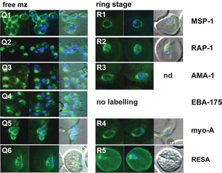
The appearance of Plasmodium falciparum merozoites and rings of the ITO4 line at different times of development. Antibodies to proteins associated with invasion related organelles or the parasite periphery (rhoptries : RAP-1; micronemes: AMA-1, EBA-175; dense granules: RESA; parasite periphery: MSP-1, myo-A) were chosen to follow the development and assembly of merozoites and rings.Margos G, Bannister LH, Dluzewski AR, Hopkins J, Williams IT, Mitchell GH. Correlation of structural development and differential expression of invasion-related molecules in schizonts of Plasmodium falciparum. Parasitology. 2004 129:273-87. Copyright Cambridge University Press Journals 2011
See original on MMP
PfTRAMP localizes to the rhoptries of P. falciparum schizonts and merozoites.(A) Co-localization of PfTRAMP with MSP1, EBA175 and Rh2b in schizonts and free merozoites by IFA. PfTRAMP (green) co-localizes with rhoptry marker PfRh2b (red) but does not co-localize with EBA175 (red) or merozoite surface marker MSP1 (red) in schizonts and merozoites. (B)Co-localization of PfTRAMP-GFP in transgenic P. falciparum 3D7 with MSP1, AMA1 and Clag3.1. PfTRAMP-GFP (green) co-localizes with rhoptry marker Clag 3.1 (red) but not with microneme markerPfAMA1 (red) or merozoite suface marker PfMSP1 (red) in schizonts and merozoites. Nuclei were stained with DNA intercalating dye DAPI.Siddiqui FA, Dhawan S, Singh S, Singh B, Gupta P, Pandey A, Mohmmed A, Gaur D, Chitnis CE. A Thrombospondin Structural Repeat Containing Rhoptry Protein from Plasmodium falciparum Mediates Erythrocyte Invasion. Cell Microbiol. 2013 15(8):1341-56
See original on MMP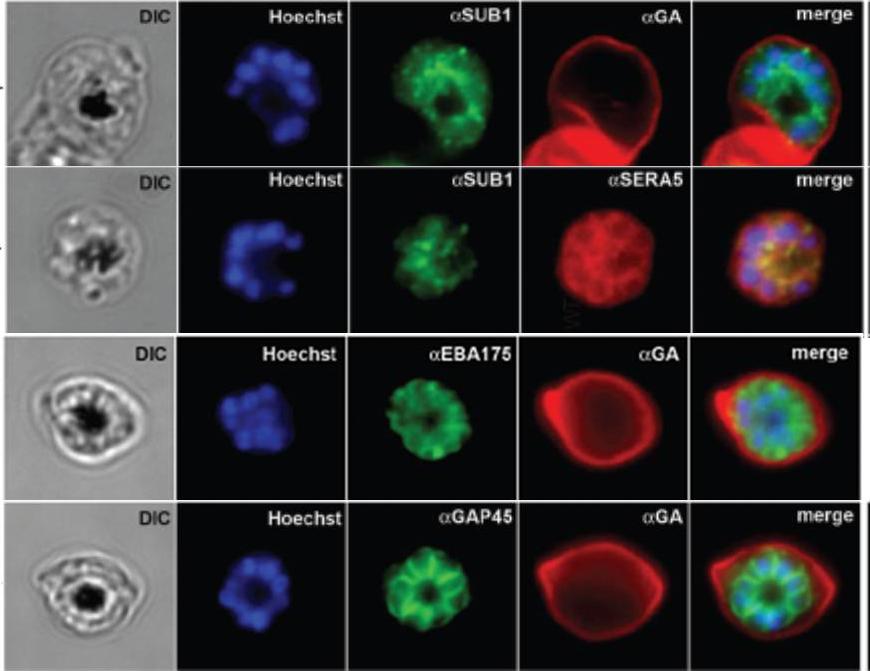
IFA of WT schizonts with glycophorin A (GA) labeled erythrocytes, localized SUB1 in an apical punctate pattern similar to SERA5. EBA-175 was detected in the apical ends of merozoites. GAP45 appeared submembrane. DIC, differential interference contrast; Hoechst = nucleic acid stain.Balu B, Maher SP, Pance A, Chauhan C, Naumov AV, Andrews RM, Ellis PD, Khan SM, Lin JW, Janse CJ, Rayner JC, Adams JH. CCR4-associated factor-1 coordinates expression of Plasmodium falciparum egress and invasion proteins. Eukaryot Cell. 2011 10(9):1257-63
See original on MMP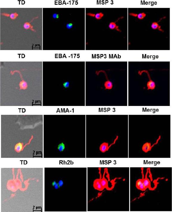
Detection of fibrillar structures in Plasmodium falciparum merozoites by immunofluorescence assay. Co-immunostaining of free merozoites with anti-MSP3 Abs along with Abs against EBA-175, AMA-1, Rh2b and MSP5. In all the slides long fibrous structure attached to free merozoite was detected by anti-MSP3 Abs. Monoclonal MSP3 antibodies also stained these fibrillar structures associated with merozoites, but other marker protein did not detect fibrillar assemblies. The long fibrils like structures attached at one end with merozoite surface were consistently seen to associate to distant erythrocytes as well as wrap around them.Imam M, Singh S, Kaushik NK, Chauhan VS. Plasmodium falciparum Merozoite Surface Protein 3: oligomerization, self-assembly and heme complex formation. J Biol Chem. 2013 289(7):3856-68
See original on MMP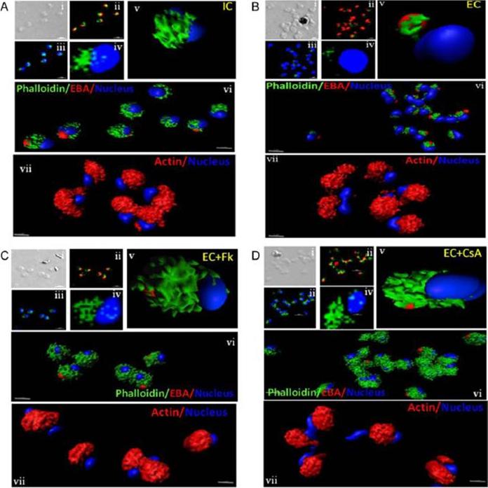
Actin polymerization status in P. falciparum merozoites. Merozoites were stained with Alexa 488-labeled phalloidin to detect polymerized F-actin (green), mouse anti EBA-175 mouse sera and Alexa Fluor 594-conjugated anti-mouse IgG goat antibodies to define location of micronemes (red) and DAPI to define location of nuclei (blue). Slides were observed under a confocal laser microscope (Nikon A1R). Bright field (i) and single slice confocal fluorescence images of merozoites (ii – iv) are shown. Three-dimensional reconstruction of confocal z stack fluorescence images of merozoites was performed using Imaris software (v –vii). Total actin (polymerized F-actin and monomeric G-actin) was detected with anti-actin rabbit sera followed by Alexa Fluor 594-conjugated anti-rabbit IgG goat antibodies (red in vii). Levels of total actin are similar in all conditions tested. Transfer of merozoites from IC to EC buffer leads to disassembly of polymerized F-actin at the apical tip of merozoites. Treatment of merozoites with FK506 and CsA prior to transfer to EC buffer leads to accumulation of polymerized actin at the apical end. Actin depolymerizing agent CytD and actin stabilizing agent JAS were used as control and result in reduced F-actin and increased F-actin at the apical tip of the merozoites respectively. Scale bar represents 1 μm. Treatment of merozoites with actin depolymerizing agents, CytD, ML-B and LA-B, resulted in increased secretion of microneme protein PfAMA-1 (a-c) whereas treatment with polymerized actin stabilizer JAS resulted in reduced secretion of PfAMA-1 (d).Singh S, More KR, Chitnis CE. Role of Calcineurin and Actin Dynamics in Regulated Secretion of Microneme Proteins in Plasmodium falciparum Merozoites during Erythrocyte Invasion. Cell Microbiol. 2013 16(1), 50–63
See original on MMP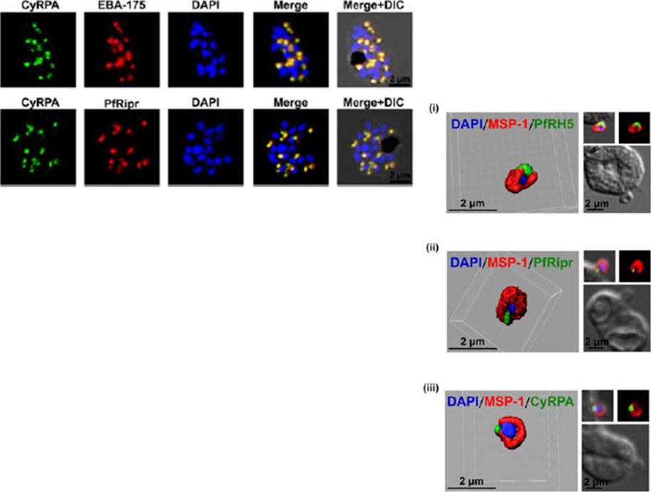
Left: Immunofluorescence microscopy detected CyRPA to colocalize with micronemal proteins (EBA-175, PfRipr). (Scale bar, 2 μm.)Right: PfRH5/PfRipr/CyRPA complex is GPI-anchored to the merozoite surface. (A) 3D reconstruction of IFA z-stacks during merozoite invasion, coimmunostained with MSP-1 detected PfRH5, PfRipr, and CyRPA at the apical end of the merozoite surface. In the 3D images, the γ settings were altered for visual representation only,Reddy KS, Amlabu E, Pandey AK, Mitra P, Chauhan VS, Gaur D. Multiprotein complex between the GPI-anchored CyRPA with PfRH5 and PfRipr is crucial for Plasmodium falciparum erythrocyte invasion. Proc Natl Acad Sci U S A. 2015 Jan 12.
See original on MMP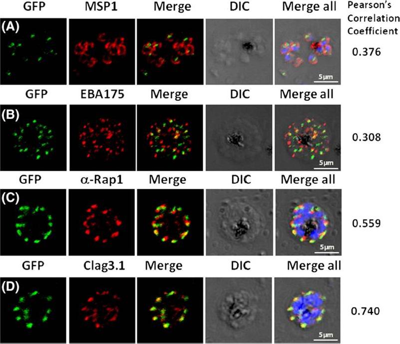
Pfμ1 co-localizeswith rhoptrymarker proteins in schizont stage parasites. Transgenic parasites expressing Pfμ1–GFPwere immunostained with antibodies specific to the Merozoite surface localized MSP1 (A), Microneme localized EBA175 (B), and Rhoptry localized RAP1 (C) and Clag3.1 (D). The parasite nuclei were stained with DAPI and slides were visualized by confocal microscopy. Representative images are shown for each antibody, together with DIC images; scale bars denote 5 μM. To quantify co-localisation, Pearson correlation coefficients of the individual stains were calculated and are shown in the right panel of each image. IFA with antibodies to rhoptry (RAP1 and Clag3.1), microneme (EBA175), and surface markers (MSP1). IFAwith anti-MSP1 antibody showed no overlap in staining betweenMSP1 and Pfμ1 (A). Similar results were seen with antibodies to EBA175 (B). Importantly, anti-RAP1 and anti-Clag3.1 showed co-localization with the Pfμ1–GFP chimeric protein (C and D), suggesting a potential role for Pfμ1 in rhoptry trafficking. Co-localization between Pfμ1 and RAP1 was first observed ~24 h post invasion in budding vesicles near the Golgi. As nuclear division commenced (32 h), Golgi multiplication occurred as well, and this resulted in apical distribution of Pfμ1 along with RAP1 in the rhoptries. Kaderi Kibria KM, Rawat K, Klinger CM, Datta G, Panchal M, Singh S, Iyer GR, Kaur I, Sharma V, Dacks JB, Mohmmed A, Malhotra P. A role for adaptor protein complex 1 in protein targeting to rhoptry organelles in Plasmodium falciparum. Biochim Biophys Acta. 2015 1853(3):699-710.
See original on MMP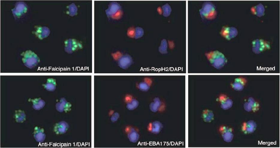
Localization of falcipain 1 (FP1) in merozoites by immunofluorescence imaging. Samples of merozoites were smeared and fixed in 1% paraformaldehyde. The slides were incubated with an antibody to falcipain 1 or with antibodies specific to the rhoptry (RopH2) and microneme (EBA-175) proteins. Nuclei were stained with 4’,6-diamidino-2-phenylindole (DAPI). The lack of colocalization indicates that falcipain 1 is found in compartments at the apical end of the merozoite that are distinct from the rhoptries and micronemes.Greenbaum DC, Baruch A, Grainger M, Bozdech Z, Medzihradszky KF, Engel J, DeRisi J, Holder AA, Bogyo M. A role for the protease falcipain 1 in host cell invasion by the human malaria parasite. Science. 2002 298(5600):2002-6.
See original on MMP
PfMyoB-GFP is located at the apical pole of the merozoite. Indirect immunofluorescence of PfMyoB-GFP (green) with various merozoite organelle markers (red), using antisera to i) EBA175 (microneme marker), ii) RAP1 (rhoptry bulb), iii) RON4 (rhoptry neck) and iv) α-tubulin. Samples were counterstained with DAPI (blue). The merged images are also shown. Rows i-iii show individual schizonts, row iv shows an individual merozoite. Scale bar: 2 μM. Antibodies to GFP produced a compact discrete dot pattern of fluorescence located at the very apical end of the MyoB-GFP parasites, near to the localisation of the apical markers EBA175, RON4, and RAP1. However, whilst in some cases the fluorescent signal partially overlapped with these markers, MyoB appeared to be in a distinct location within the cell, anterior to the microneme marker, the rhoptry bulb, and even to the rhoptry neck.Yusuf NA, Green JL, Wall RJ, Knuepfer E, Moon RW, Schulte-Huxel C, Stanway RR, Martin SR, Howell SA, Douse CH, Cota E, Tate EW, Tewari R, Holder AA. The Plasmodium Class XIV Myosin, MyoB has a Distinct Subcellular Location in Invasive and Motile Stages of the Malaria Parasite, and an Unusual Light Chain. J Biol Chem. 2015 Mar 23.
See original on MMP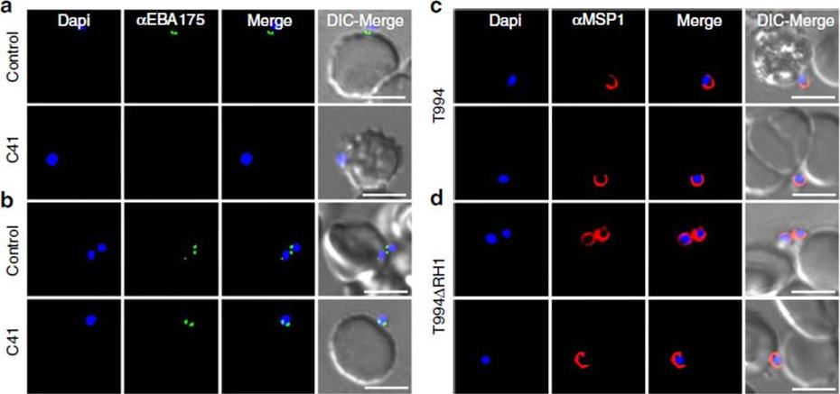
Effect of anti-PfRH1 inhibitory mAbs on merozoite surface expression of EBA175 during invasion. (a–d) Surface expression of EBA175 and MSP1 on T994 and T994DRH1 was detected by immunofluorescence assays. Isolated T994 (a,c) or T994DRH1 (b,d) merozoites were directly incubated with erythrocytes containing 4 mM Cyto D in the absence (Control) and presence (C41) of C41 (0.2mgml1) before analysis of expression of EBA175 and MSP1 at the junction. Both differential interference contrast (DIC) and fluorescence images were captured. Nuclear DNA was counterstained with DAPI (blue). EBA175 is shown in green, whereas MSP1 is shown in red. Fluorescence images, merged fluorescence images (DAPI and green or DAPI with red) and merged fluorescence images with DIC images are shown. Scale bars=10 mM. Antibody C41 blocks surface expression of EBA175 in T994 (a lower panel), whereas it has no effect on T994DRH1 parasites (b lower panel). In the absence of C41, EBA175 is expressed on the surface of both T994 and T994DRH1 (upper panels a,b). Ab has no effect on the detection of MSP1 on the surface of merozoites (c,d).Gao X, Gunalan K, Yap SS, Preiser PR. Triggers of key calcium signals during erythrocyte invasion by Plasmodium falciparum. Nat Commun. 2013;4:2862.
See original on MMP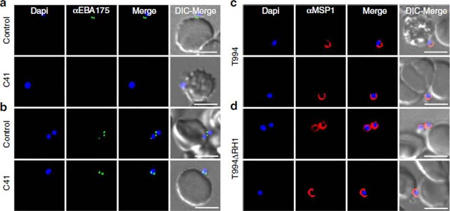
Effect of anti-PfRH1 inhibitory mAbs on merozoite surface expression of EBA175 during invasion. Isolated T994 (a,c) were directly incubated with erythrocytes containing 4 mM Cyto D in the absence (Control) and presence (C41) of C41 (0.2mgml-1) before analysis of expression of microneme protein EBA175 and merozoite surface marker MSP1 at the junction. Both differential interference contrast (DIC) and fluorescence images were captured. Nuclear DNA was counterstained with DAPI (blue). EBA175 is shown in green, whereas MSP1 is shown in red. Fluorescence images, merged fluorescence images (DAPI and green or DAPI with red) and merged fluorescence images with DIC images are shown. Scale bars=10 mm.Gao X, Gunalan K, Yap SS, Preiser PR. Triggers of key calcium signals during erythrocyte invasion by Plasmodium falciparum. Nat Commun. 2013;4:2862. PMID:
See original on MMP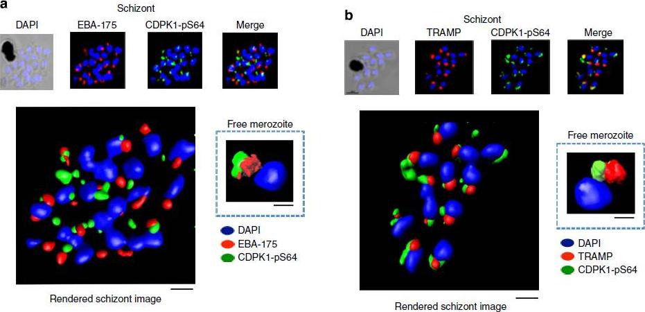
PfCDPK1 phosphorylated at serine-64 is associated with apical parasite structures. (a) A schizont stage parasite stained with DAPI to reveal the nuclei (blue), an antibody to PfEBA-175 to reveal the micronemes (red) and the phospho-specific CDPK1-pS64 antibody (green). A merge of images from all three stains is shown on the far right; this is a representative image from at least three experiments. Also shown is a rendered image where deconvoluted z stacks were reconstructed in 3D, with interpolation. The inset shows a rendered image of a free merozoite from the same preparation . (b) The same as a, but instead of probing with an anti-EBA-175 antibody the preparation was probed with an antibody to the rhoptrymarker PfTRAMP . Scale bars, a-1 mm (insert-0.5 mm), b-1 mm (insert-0.5 mm).Alam MM, Solyakov L, Bottrill AR, Flueck C, Siddiqui FA, Singh S, Mistry S, Viskaduraki M, Lee K, Hopp CS, Chitnis CE, Doerig C, Moon RW, Green JL, Holder AA, Baker DA, Tobin AB. Phosphoproteomics reveals malaria parasite Protein Kinase G as a signalling hub regulating egress and invasion. Nat Commun. 2015 6:7285
See original on MMP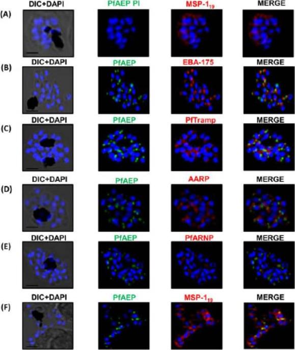
PfAEP localization in schizont stages analyzed by confocal microscopy. Subcellular localization of PfAEP was studied by co-staining with antibodies against micronemal protein EBA-175 (B), rhoptry bulb protein PTRAMP (C), and rhoptry neck,apical asparagine rich protein (AARP) (D), apical rhoptry neck protein (ARNP) (E), and merozoite surface protein (MSP) (F).As a control, pre-immune serum of PfAEP was checked and no staining was observed in schizonts (A). Mature schizonts were costained with anti-PfAEP (green) and anti-EBA-175, TRAMP, AARP, ARNP, MSP-119 antibodies (red). The nuclei of schizonts were stained with DAPI (blue) and visualized by Nikon N-SIM confocal microscope. All apical marker proteins and PfAEP showed punctate staining in schizonts distinct from DAPI. Co-staining of PfAEP with surface marker MSP-119 showed PfARNP localized at the apical tip with surface of merozoites stained by MSP-119. PfAEP staining did not merge with either markers of microneme or rhoptry indicating that PfAEP is neither a resident of micronemes or rhoptry ofmerozoites. Scale bar 2 mm.Hans N, Relan U, Dubey N, Gaur D, Chauhan VS. Identification and localization of a Novel Invasin of Plasmodium falciparum. Mol Biochem Parasitol. 2015 Sep 29. [Epub ahead of print]
See original on MMP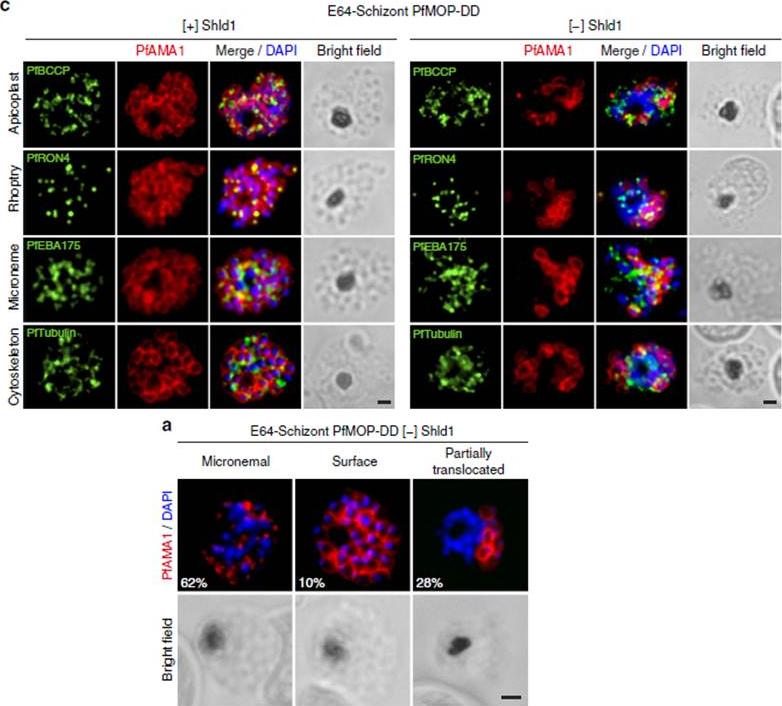
PfAMA1 translocation is aberrant in schizonts with PfMOP-knockdown. (a) Schizonts from [+]/[-] Shld1 PfMOP-DD parasites were E64-treated, fixed, probed with anti-PfAMA1 and scored as micronemal (M), partially translocated (PT) or surface (S), representative [+] Shld1 parasite IFAs shown. (c) Synchronized schizont stage (40–44 h) parasites, maintained with 250nM (left panel) or 0 nM (right panel) Shld1, were incubated 6 h in presence of 10 mM E64, methanol-fixed, permeabilized, and stained using antibodies against PfRON4, PfRhopH3, PfEBA175 and PfTubulin. Staining for these markers was similar in [+] and [-] Shld1 conditions. PfAMA1 staining was used to identify E64-treated schizonts that were sufficiently mature (that is, surface staining or partially translocated staining, but not micronemal staining, scale bar, 1 mm).Absalon S, Robbins JA, Dvorin JD. An essential malaria protein defines the architecture of blood-stage and transmission-stage parasites. Nat Commun. 2016 7:11449.
See original on MMP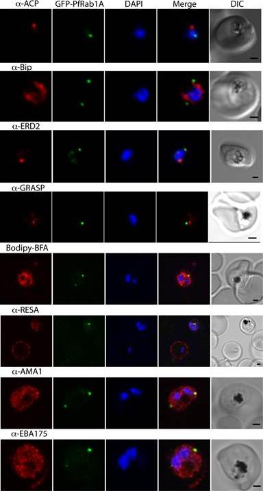
GFP-PfRab1A fluorescence does not co-localize with ER/Golgi, dense granules or microneme markers. GFP-PfRab1A fluorescence is distinct from the localization of markers for the apicoplast (ACP), the ER (Bip), the Golgi (ERD2 and GRASP), as well as from staining of the ER/Golgi with Bodipy BFA. GFP-Rab1A fluorescence is also distinct from the localization of markers for dense granules (RESA) or micronemes (AMA1 and EBA175).Morse D, Webster W, Kalanon M, Langsley G, McFadden GI. Plasmodium falciparum Rab1A Localizes to Rhoptries in Schizonts. PLoS One. 2016 Jun 27;11(6):e0158174
See original on MMP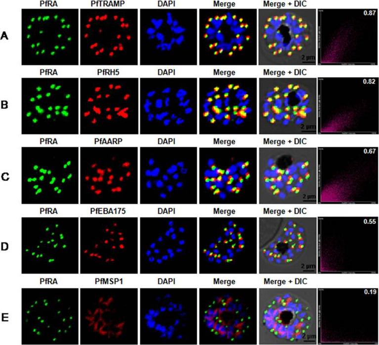
PfRA localizes to the rhoptries of P. falciparum schizonts and merozoites. Co-localization of PfRA with (A) PfTRAMP, (B) PfRH5, (C) PfAARP, (D) PfEBA-175 and (E) MSP-1 in schizont stage parasites by confocal immunoflourescence microscopy. PfRA (green) co-localizes with rhoptry bulb protein PfTRAMP(red) and PfRH5 (red) but does not co-localize with the micronemal protein PfEBA-175 (red) and merozoite surface protein MSP-1 in schizonts. PfRA (green) showed partial co-localization with rhoptry neck protein PfAARP (red). The co-localization of PfRA was quantitatively analyzed and the Pearson correlation co-efficientwith PfTRAMP (0.87) and PfRH5 (0.82) suggested that the two proteins were co-localized with PfRA. The coefficients of PfRA with PfAARP, PfEBA-175 and MSP1 were lower than 0.7 indicating that the three proteins were not co-localized with PfRA. Nuclei were stained with DNA intercalating dye DAPI. (Scale bar, 2 μm.)Anand G, Reddy KS, Pandey AK, Mian SY, Singh H, Mittal SA, Amlabu E, Bassat Q, Mayor A, Chauhan VS, Gaur D. A novel Plasmodium falciparum rhoptry associated adhesin mediates erythrocyte invasion through the sialic-acid dependent pathway. Sci Rep. 2016 6:29185.
See original on MMP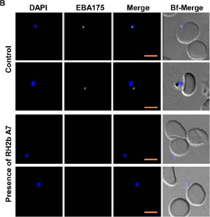
EBA175 surface expression is inhibited by mAb A7. (B) Representative images indicate the positivity of merozoites to EBA175 surface expression upon incubation with PfRH2b A7 or without A7 (Control). Merozoites nuclei shown in blue (DAPI) and EBA175 surface staining indicated in green color. Scale bar indicates 2 μm. Aniweh Y, Gao X, Gunalan K, Preiser PR. PfRH2b Specific Monoclonal Antibodies Inhibit Merozoite Invasion. Mol Microbiol. 2016 Jul 20.
See original on MMP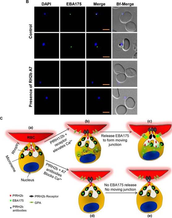
B. Representative images indicate the positivity of merozoites to EBA175 surface expression upon incubation with PfRH2b A7 or without A7 (Control). Merozoites nuclei shown in blue (DAPI) and EBA175 surface staining indicated in green color. Scale bar indicates 2 mm.C. Schematic of the effect of PfRH2b A7 on merozoite Ca2+ signalling and invasion. After the merozoite initially attaches to the RBC and reorient itself, PfRH2b is released from rhoptry neck aiding in the sensing of the RBC by engaging in apical end interaction with the surface of the RBC (a). The interaction between PfRH2b and its receptor triggers the increase of intracellular Ca2+ of the merozoite (b). The increase in cytosolic Ca2+ levels lead toa release of microneme proteins such as EBA175 to bind to its receptor glycophorin A (GPA). This therefore triggers the recruitment of other members leading to moving/tight junction formation (c). In the presence of PfRH2b A7 mAb, interactions between PfRH2b and its receptor is abrogated thereby affecting the intracellular Ca2+ increase (d) necessary for the release of EBA175. This, therefore, hinders recruitment of other proteins, such as AMA1,needed for merozoite tight junction formation. Hence no tight or moving junction is formed leading to invasion blocking.Aniweh Y, Gao X, Gunalan K, Preiser PR. PfRH2b Specific Monoclonal Antibodies Inhibit Merozoite Invasion. Mol Microbiol. 2016 Jul 20.
See original on MMP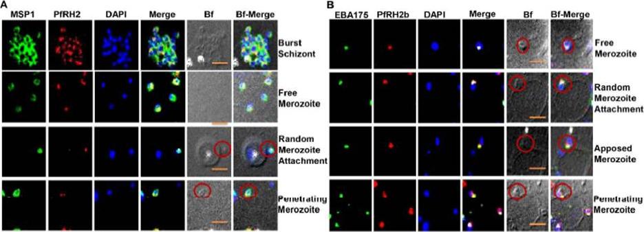
PfRH2b is associated with the moving junction during merozoite invasion. The process of merozoite invasion was closely monitored to locate the position of PfRH2b of burst schizont, free merozoites, random merozoite attachment, apposed merozoite and penetrating merozoite. (A) Merozoites were stained for MSP1 (MSP1, green) and PfRH2 (PfRH2, red). PfRH2 apical punctuate staining was observed in the free and attaching merozoites while in invading merozoites two punctuate staining were observed at the anchorage of the moving junction. The presence of PfRH2b (PfRH2b, red) at the moving junction was further confirmedwith EBA175 (B, green). Merozoites of focus are shown with red circles. In the merged images, areas of overlap between the red and the green signals are shown in yellow. DAPI (blue) stained for nuclei.Aniweh Y, Gao X, Gunalan K, Preiser PR. PfRH2b Specific Monoclonal Antibodies Inhibit Merozoite Invasion. Mol Microbiol. 2016 Jul 20. .
See original on MMP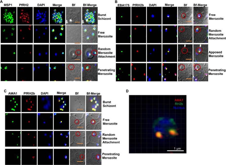
PfRH2b is associated with the moving junction during merozoite invasion. The process of merozoite invasion was closely monitored to locate the position of PfRH2b of burst schizont, free merozoites, random merozoite attachment, apposed merozoite and penetrating merozoite. A. Merozoites were stained for MSP1 (MSP1, green) and PfRH2 (PfRH2, red). PfRH2 apical punctuate staining was observed in the free and attaching merozoites while in invading merozoites two punctuate staining were observed at the anchorage of the moving junction. The presence of PfRH2b (PfRH2b, red) at the moving junction was further confirmed with EBA175 (B, green) and AMA1 (C, green) as common markers with punctuate staining. Merozoites of focus are shown with red circles. The bright field images labelled as Bf. Scale bar indicates 2 lm. D. A slice of a three-dimensional structured illumination microscopy (3D-SIM) on merozoite at the junction further confirmed the punctuated pattern of PfRH2b (green) to be in close proximity to AMA1 (red). Scale bar indicates 1 lm. In the merged images, areas of overlap between the red and the green signals are shown in yellow. DAPI (blue) stained for nuclei.Aniweh Y, Gao X, Gunalan K, Preiser PR. PfRH2b specific monoclonal antibodies inhibit merozoite invasion. Mol Microbiol. 2016 Nov;102(3):386-404.
See original on MMP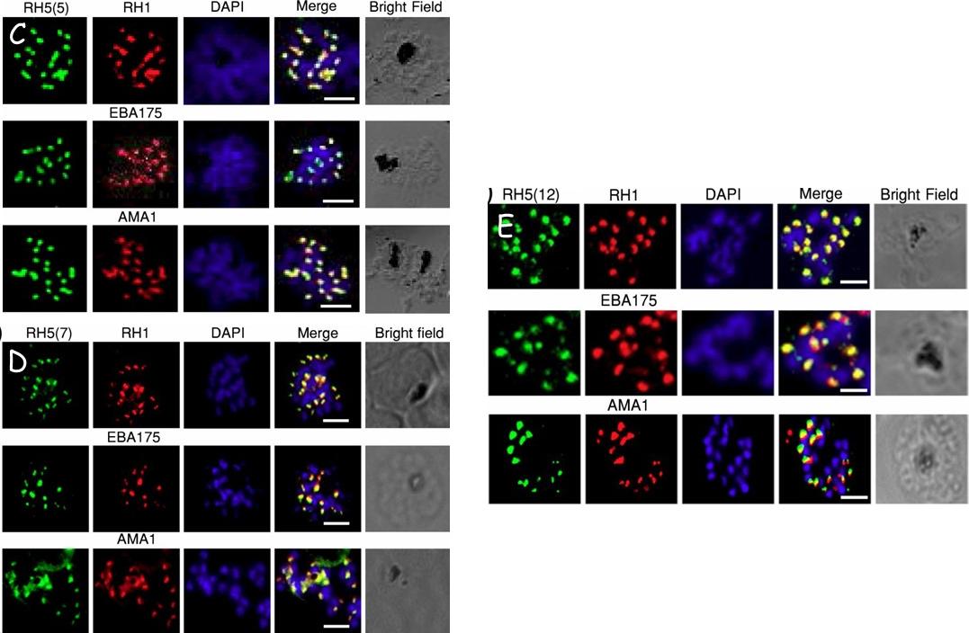
Validation of RH5 mAbs by Immunofluroscence Microscopy. Smears of 3D7 late stage schizont were co- stained with RH5(5) (c) (7) (d) and (12) (e) (first column, green) and antibodies against other invasion markers RH1, EBA175 and AMA1 respectively (second column, red). Parasite nuclei were stained with DAPI (blue). In the merged images, areas of overlap between the red and the green signals are shown in yellow. Bright field indicates the outline of the schizont. Scale bar =2μm.Aniweh Y, Gao X, Hao P, Meng W, Lai SK, Gunalan K, Chu TT, Sinha A, Lescar J, Chandramohanadas R, Li HY, Sze SK, Preiser PR. RH5-Basigin interaction induceschanges in the cytoskeleton of the host RBC. Cell Microbiol. 2017 Apr 13 [Epub ahead of print] PMID: 28409866
See original on MMPMore information
| PlasmoDB | PF3D7_0731500 |
| GeneDB | PF3D7_0731500 |
| Malaria Metabolic Pathways | Localisation images Pathways mapped to |
| Previous ID(s) | MAL7P1.176, PF07_0128 |
| Orthologs | |
| Google Scholar | Search for all mentions of this gene |