PF3D7_0731100 EMP1-trafficking protein (PTP2)
Disruptability [+]
| Species | Disruptability | Reference | Submitter |
|---|---|---|---|
| P. falciparum 3D7 |
Possible |
18614010 | Theo Sanderson, Wellcome Trust Sanger Institute |
| P. falciparum 3D7 |
Possible |
USF piggyBac screen (Insert. mut.) | USF PiggyBac Screen |
Mutant phenotypes [+]
None reported yet. Please press the '+' button above to add one.Imaging data (from Malaria Metabolic Pathways)
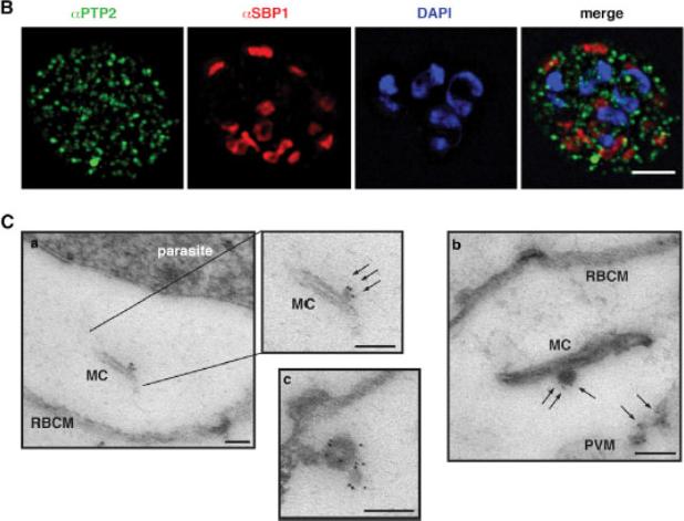
Localization of PfPTP2 in CS2idhfrPTP2/HA-infected RBCs. (B) First panel, PfPTP2 (green); second panel, SBP1 (Maurer’s cleft marker; red); third panel, DAPI (blue); fourth panel, all panels merged. (C) Immuno-EM of CS2idhfrPTP2/HA-infected RBCs after treatment with equinotoxin II. MC, Maurer’s cleft; RBCM, red blood cell plasma membrane; PVM, parasitophorous vacuole membrane. a, the side panel shows higher magnification of Maurer’s cleft; arrows point to budding vesicle where PfPTP2 is localized. b, arrows point to PfPTP2 on budding vesicle and membrane material. c, an example of budding vesicle with PfPTP2 localization. PfPTP2-labeled material in host cell cytoplasm was derived from Maurer’s clefts .Regev-Rudzki N, Wilson DW, Carvalho TG, Sisquella X, Coleman BM, Rug M, Bursac D, Angrisano F, Gee M, Hill AF, Baum J, Cowman AF. Cell-Cell Communication between Malaria-Infected Red Blood Cells via Exosome-like Vesicles. Cell. 2013 May 23;153(5):1120-33.
See original on MMP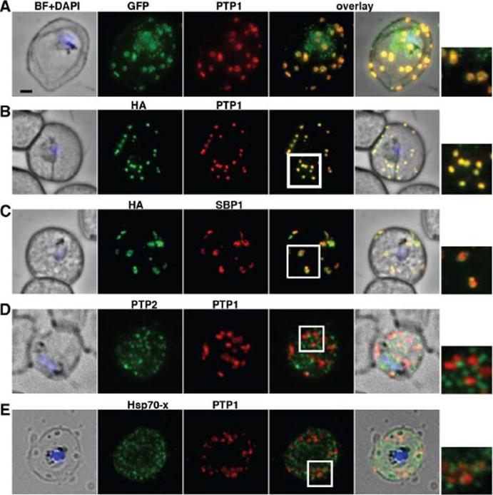
PfPTP1 is localised on Maurer’s clefts in P. falciparum-infected RBCs. (A) Immunofluorescence assay on CS2/PfPTP1-GFP cell line: the GFP signal (green) colocalises with that of the endogenous protein (PfPTP1; red); inset: enlargement of colocalisation pattern. (B) Immuno-fluorescence assay on CS2/PfPTP1-HA cell line: the HA signal (green) colocalises with the α-PfPTP1 signal (red); inset: enlargement of colocalisation pattern. (C) Immunofluorescence assay on CS2/PfPTP1-HA cell line: the HA-signal (green) colocalises partially with the MC resident protein SBP1 (red); inset: enlargement of colocalisation pattern. (D) Immunofluorescence assay on CS2 parental cell line: the PfPTP2-signal (green) does not overlap with the PfPTP1-signal (red); inset: enlargement of area where MC (PfPTP1) and electron dense vesicles (PfPTP2) are in close proximity. (E) Immunofluorescence assay on CS2 parental cell line: the PfHsp70-x-signal (green) colocalises partially with the PfPTP1- signal (red); inset: enlargement of partial colocalisation on J-Dots (PfHsp70-x). Scale bar: 1μm; same size of ROI in each image. Rug M, Cyrklaff M, Mikkonen A, Lemgruber L, Kuelzer S, Sanchez CP, Thompson J, Hanssen E, O'Neill M, Langer C, Lanzer M, Frischknecht F, Maier AG, Cowman AF. Export of virulence proteins by malaria-infected erythrocytes involves remodelling of host actin cytoskeleton. Blood. 2014 Aug 19 [Epub ahead of print]
See original on MMP
Localisation of PHIST:GFP proteins. The left- and right-hand images show GFP localisation and a phase contrast image, respectively. White filled arrows: peripheral GFP puncta; White unfilled arrow: GFP puncta in host erythocyte cytosol. Scale bar, 2 mm. GFP-tagged PF3D7_0401800, PF3D7_0424600, PF3D7_0532400, PF3D7_1102500 and PF3D7_1476200 were all exported to the host cell and displayed a striking localisation at the edge of the host erythrocyte (1A–E), PF3D7 1252700 was clearly peripheral in the host erythrocyte (F). PF3D7_0201600 (G) shows weak accumulation at the erythrocyte periphery, but the majority of the protein was localised in the RBC cytosol. PF3D7_0936600 was localised in the erythrocyte cytosol, not at the host cell periphery (H).Tarr SJ, Moon RW, Hardege I, Osborne AR. A conserved domain targets exported PHISTb family proteins to the periphery of Plasmodium infected erythrocytes. Mol Biochem Parasitol. 2014 196(1):29-40 PMID: 25106850
See original on MMP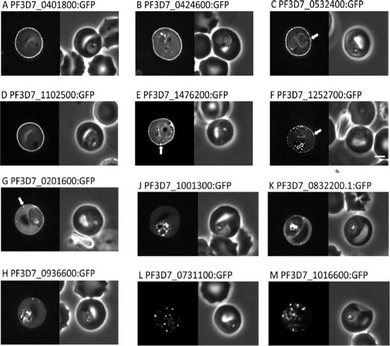
Localisation of PHIST:GFP proteins. The left- and right-hand images show GFP localisation and a phase contrast image, respectively. White filled arrows: peripheral GFP puncta; White unfilled arrow: GFP puncta in host erythocyte cytosol. Scale bar, 2 mm. GFP-tagged PF3D7_0401800, PF3D7_0424600, PF3D7_0532400, PF3D7_1102500 and PF3D7_1476200 were all exported to the host cell and displayed a striking localisation at the edge of the host erythrocyte (1A–E), PF3D7 1252700 was clearly peripheral in the host erythrocyte (F). PF3D7_0201600 (G) shows weak accumulation at the erythrocyte periphery, but the majority of the protein was localised in the RBC cytosol. PF3D7_0936600 was localised in the erythrocyte cytosol, not at the host cell periphery (H).Tarr SJ, Moon RW, Hardege I, Osborne AR. A conserved domain targets exported PHISTb family proteins to the periphery of Plasmodium infected erythrocytes. Mol Biochem Parasitol. 2014 196(1):29-40
See original on MMPMore information
| PlasmoDB | PF3D7_0731100 |
| GeneDB | PF3D7_0731100 |
| Malaria Metabolic Pathways | Localisation images Pathways mapped to |
| Previous ID(s) | MAL7P1.172 |
| Orthologs | |
| Google Scholar | Search for all mentions of this gene |