PF3D7_0714400 calmodulin, putative
Disruptability [+]
| Species | Disruptability | Reference | Submitter |
|---|---|---|---|
| P. falciparum 3D7 |
Possible |
USF piggyBac screen (Insert. mut.) | USF PiggyBac Screen |
Mutant phenotypes [+]
None reported yet. Please press the '+' button above to add one.Imaging data (from Malaria Metabolic Pathways)
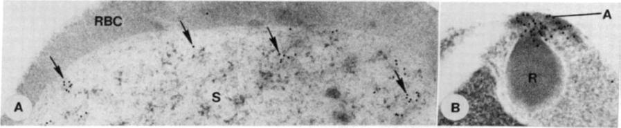
Immunoelectron micrographs. (A) Section of an early schizont of P.falciparum (Brazilian strain 7G8, drug sensitive) incubated with anti-CaM antibody and protein A-gold. Gold particles (arrows) are scattered diffusely over the schizont (S) cytoplasm. Little label is associated with the host erythrocyte (RBC). (x43,000.) (B) Section of merozoite. Gold particles indicate the location of CaM at the apical end (A) and the ductule of rhoptries (R). (x52,000.)Scheibel LW, Colombani PM, Hess AD, Aikawa M, Atkinson CT, Milhous WK. Calcium and calmodulin antagonists inhibit human malaria parasites (Plasmodium falciparum): implications for drug design. Proc Natl Acad Sci U S A. 1987 84:7310-4.
See original on MMP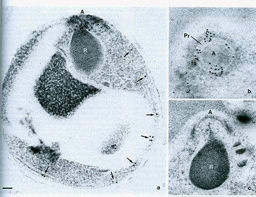
Ultrathin sections of merozoites were incubated with sheep Abs to calmodulin from bovine testis (these Abs are specific for all calmodulins found in all eukaryotic cells; It is not clear if the Ab reacted with all homologs of calmodulin or specifically with some of them) and then with protein-A gold. a. an are of intense immunoreactivity is located on the merozoite apical end. Some label was also noted immediately under the plasma membrane (arrows). b, e) Tangential section of the apical end of maturing merozoites, the calmodulin labeling is mostly on the polar rings.. A – apical end; R – rhoptry; Pr – polar ring. Bar= 0.1 mm.Matsumoto Y, Perry G, Scheibel LW, Aikawa M. Role of calmodulin in Plasmodium falciparum: implications for erythrocyte invasion by the merozoite. Eur J Cell Biol. 198745:36-43. Copyright Elsevier 2009.
See original on MMP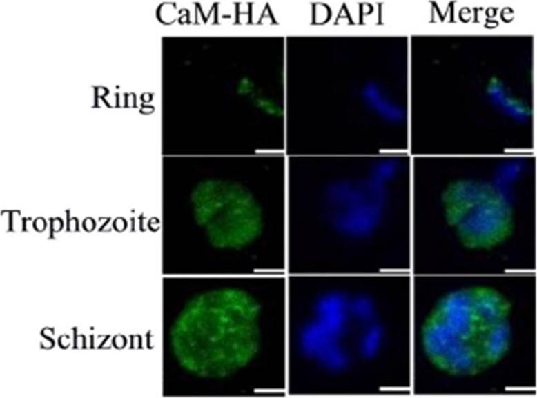
Detection of CaM via immunofluorescence in intraerythrocytic stages with anti-HA and anti-IgG rabbit Alexa 488 (green). Nucleus was stained with DAPI (blue). Confocal images are shown as the maximum projection of stacks. Scale bars are of 2 μm. The immunofluorescence in CaM-Str-HA parasites indicated that the distribution of CaM is consistent with that of a cytosolic protein.Budu A, Gomes MM, Melo PM, El Chamy Maluf S, Bagnaresi P, de Azevedo MF, Carmona AK, Gazarini ML. Calmidazolium evokes high calcium fluctuations in Plasmodium falciparum. Cell Signal. 2015 Dec 9. [Epub ahead of print]
See original on MMPMore information
| PlasmoDB | PF3D7_0714400 |
| GeneDB | PF3D7_0714400 |
| Malaria Metabolic Pathways | Localisation images Pathways mapped to |
| Previous ID(s) | MAL7P1.69 |
| Orthologs | PBANKA_1421000 , PCHAS_1422800 , PKNH_1423300 , PVP01_1423500 , PVX_122947 , PY17X_1422700 |
| Google Scholar | Search for all mentions of this gene |