PF3D7_0631900 stevor
Disruptability [+]
| Species | Disruptability | Reference | Submitter |
|---|---|---|---|
| P. falciparum 3D7 |
Refractory |
USF piggyBac screen (Insert. mut.) | USF PiggyBac Screen |
| P. falciparum 3D7 |
Possible |
31363031 | Jan Stephan Wichers, |
Mutant phenotypes [+]
| Species | Stage | Phenotype | Reference | Submitter |
|---|---|---|---|---|
| P. falciparum 3D7 | Asexual |
No difference |
31363031 | Jan Stephan Wichers, |
Imaging data (from Malaria Metabolic Pathways)

Immunolocalization of epitope-tagged proteins. (A) Schematic representation of the SFM (Stevor-FLAG-myc) and 2TMFM (Pfmc-2TM-FLAG-myc) recombinant proteins. For the respective genes, two FLAG epitopes (F) were placed between the 2TM domains and three myc epitopes (M) were inserted at the carboxy terminus. The lengths, in amino acids, of the chimeric proteins are indicated at the bottom. The restriction sites specific for the insertion of tags are indicated. (B) Immunofluorescence assays (IFA) showing the localization of SFM and 2TMFM in the transformed lines, pHL-SFM and pHL-2TMFM. IFA studies were performed on air-dried P. falciparum transformed parasites. Infected erythrocytes were stained with FITC-conjugated anti-c-myc mAb and with anti-FLAG polyclonal antibodies followed by goat anti-rabbit Alexa 594-conjugated IgG. IFA microscopy studies using anti-c-myc and anti-FLAG antibodies showed that the tagged proteins were efficiently expressed within the cytoplasm of infected erythrocytes (B); and both antibodies gave staining patterns that likely correspond to Maurer’s cleft localization.Lavazec C, Sanyal S, Templeton TJ. Hypervariability within the Rifin, Stevor and Pfmc-2TM superfamilies in Plasmodium falciparum. Nucleic Acids Res. 2006;34:6696-707.
See original on MMP
Kinetics of export of SOLCAD (partial STEVOR sequence attached to PEXEL) in various P. falciparum-infected erythrocytes. P. falciparum-infected erythrocytes were taken from culture at the time points indicated after the addition of the anti-aggregation ligand and immediately analyzed by live cell confocal fluorescence microscopy. Tightly synchronized cultures at the trophozoite-stage (18–22 h post invasion) were used. Images shown are representative examples for the export phenotypes.Kilian N, Srismith S, Dittmer M, Ouermi D, Bisseye C, Simpore J, Cyrklaff M, Sanchez CP, Lanzer M. Hemoglobin S and C affect protein export in Plasmodium falciparum-infected erythrocytes. Biol Open. 2015 4(3):400-10.
See original on MMP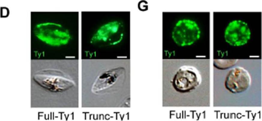
Schematic representation of STEVOR recombinant proteins overexpressed in the Full-Ty1 (PFF1550w stevor gene) and in the Trunc-Ty1 (Ty1-tagged copy of PFF1550w truncated for the cytoplasmic domain) lines. D, G. Immunofluorescence analysis of stage III GIE (D) and asexual stages (G) from the Full-Ty1 and the Trunc-Ty1 lines. Infected erythrocytes were stained with anti-Ty1 antibodies followed by anti-mouse Alexa 488-conjugated IgG. Pictures were taken under identical exposure conditions. The bars represent 2 μm.Naissant B, Dupuy F, Duffier Y, Lorthiois A, Duez J, Scholz J, Buffet P, Merckx A, Bachmann A, Lavazec C. Plasmodium falciparum STEVOR phosphorylation regulates host erythrocyte deformability enabling malaria parasite transmission. Blood. 2016 May 2. pii: blood-2016-01-690776. [Epub ahead of print] PMID: 27136945
See original on MMPMore information
| PlasmoDB | PF3D7_0631900 |
| GeneDB | PF3D7_0631900 |
| Malaria Metabolic Pathways | Localisation images Pathways mapped to |
| Previous ID(s) | 2270.t00314, MAL6P1.10, PFF1550w |
| Orthologs | |
| Google Scholar | Search for all mentions of this gene |