PF3D7_0620400 merozoite surface protein 10 (MSP10)
Mutant phenotypes [+]
None reported yet. Please press the '+' button above to add one.Imaging data (from Malaria Metabolic Pathways)
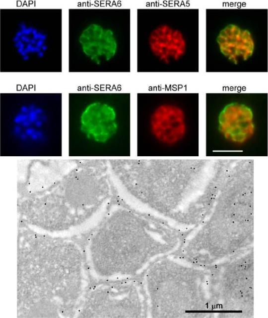
SERA6 is a soluble parasitophorous vacuole (PV) protein which may partially interact with the PVM. Upper panel: IFA demonstrates colocalization of the anti-SERA6 and anti-SERA5 signal in mature schizonts, except that the SERA6 signal additionally shows an association with the outer confines of the intracellular parasite, probably corresponding to the PVM. Scale bar, 5 μm. Lower panel: Immuno-electron microscopic localization of SERA6 in a P. falciparum schizont, using the anti-S6C1 antibodies labelled with 10 nm immunogold. The majority of the signal is clearly associated with the peripheral space between intracellular merozoites, consistent with a PV localization. Magnification, x 15000.Ruecker A, Shea M, Hackett F, Suarez C, Hirst EM, Milutinovic K, Withers-Martinez C, Blackman MJ. Proteolytic activation of the essential parasitophorous vacuole cysteine protease SERA6 accompanies malaria parasite egress from its host erythrocyte. J Biol Chem. 2012287(45):37949-63.
See original on MMP
The genes for P12 and P41 can be disrupted and are therefore not essential for parasite growth. Immunofluorescence microscopy of Dp12 and Dp41 and parental 3D7 parasites probed with rabbit anti-P12 and anti-P41 IgGs in addition to the merozoite surface marker MSP1 mAb indicate absence of protein expression in the mutants aside from minor cross-reactivity.Taechalertpaisarn T, Crosnier C, Bartholdson SJ, Hodder AN, Thompson J, Bustamante LY, Wilson DW, Sanders PR, Wright GJ, Rayner JC, Cowman AF, Gilson PR, Crabb BS. Biochemical and Functional Analysis of Two Plasmodium falciparum Blood-Stage 6-Cys Proteins: P12 and P41. PLoS One. 2012;7(7):e41937.
See original on MMP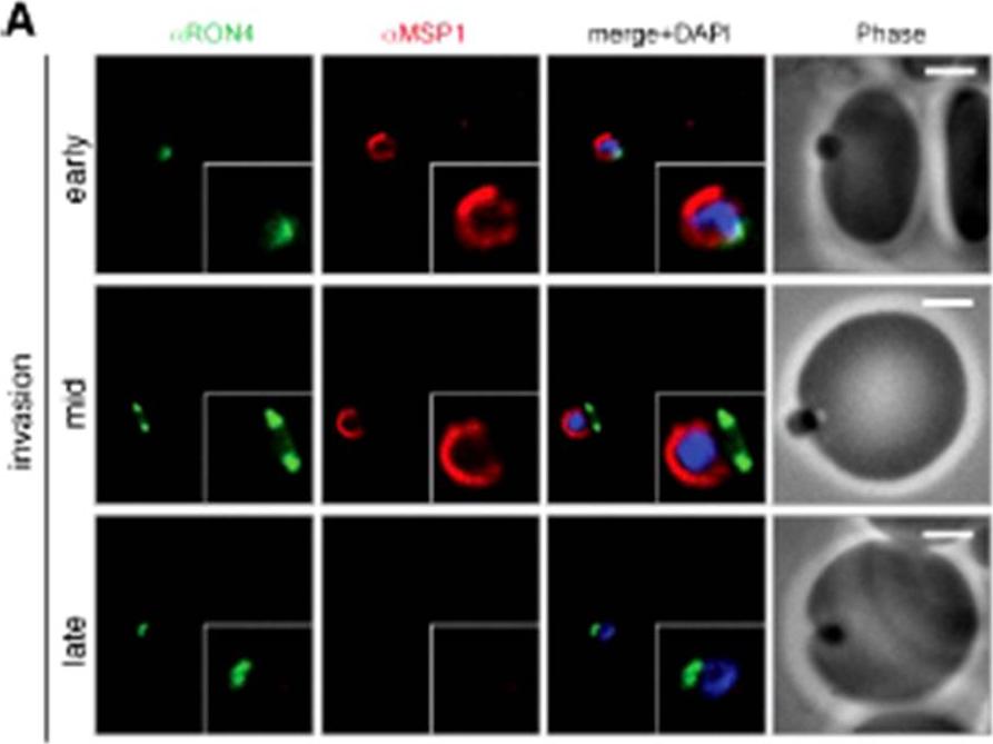
Wide field IFA time course of invasion using A) anti-MSP1/PfRON4. Scale bar = 2.0 μm. Before commencement of invasion, no loss of MSP1 labeling was apparent (MSP1 early). Loss of MSP1 tracked the exterior of invading merozoites concurrent with different stages of passage through the tight (MSP1 mid). The lack of contiguity between shed MSP1 and PfSUB2 sheddase, each in comparison with PfRON4 suggests that MSP1 may be processed by PfSUB2 early in invasion but shed only during passage through the junction.Riglar DT, Richard D, Wilson DW, Boyle MJ, Dekiwadia C, Turnbull L, Angrisano F, Marapana DS, Rogers KL, Whitchurch CB, Beeson JG, Cowman AF, Ralph SA, Baum J. Super-resolution dissection of coordinated events during malaria parasite invasion of the human erythrocyte. Cell Host Microbe. 2011 9:9-20.
See original on MMP
Two-color indirect immunofluorescence confocal microscopy of fixed smears of P. falciparum 3D7 merozoites. Panels A and E represent images generated by differential interference contrast (DIC) microscopy of the fixed smears incubated with (B) anti-MSP10B rabbit antiserum (red), (C) 1E1, a mAb reactive with MSP119 (green), (D) merge of micrographs B and C, (F) anti-MSP10B rabbit antiserum (red), (G) anti-RAMA mouse antiserum (green), (H) merge of micrographs F and G. The protein partitions in the detergent-enriched phase after Triton X-114 fractionation and is localized to the surfaces of trophozoites, schizonts and free merozoites in an apical distribution.Black CG, Wang L, Wu T, Coppel RL. Apical location of a novel EGF-like domain-containing protein of Plasmodium falciparum. Mol Biochem Parasitol. 2003 127:59-68. Copyright Elsevier 2009. PMID:
See original on MMP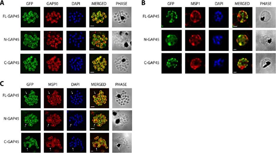
Immunofluorescent staining of GFP-tagged GAP45 variants with GAP50 in late stage schizonts. (A) Parasites were fixed and the location of GFP (green) and GAP50 (red), examined. FL-GAP45, C-GAP45 and N-GAP45 all appear to colocalise with GAP50 in segmented schizonts. (B) Staining with anti-MSP1 antibody (red) colocalises with only N-GFP in developing schizonts. FL-GAP45 AND C-GAP45 are present in distinctive ring-shaped structures at the periphery of the developing schizont. (C) In mature schizonts MSP1 (red) is present on membranes surrounding the residual body. N-GAP45 is also present on the residual body, but FL-GAP45 and C-GAP45 are absent. The white arrow in all images indicates the residual body. These data are consistent with a plasma membrane location for N-GAP45 and an inner membrane complex location for FL-GAP45. In all cases, parasite nuclei were stained with DAPI (blue); merged images are also shown. Scale bar is 2 mm.Immunofluorescent staining of GFP-tagged GAP45 variants with GAP50 in late stage schizonts. (A) Parasites were fixed and the location of GFP (green) and GAP50 (red), examined. FL-GAP45, C-GAP45 and N-GAP45 all appear to colocalise with GAP50 in segmented schizonts. (B) Staining with anti-MSP1 antibody (red) colocalises with only N-GFP in developing schizonts. FL-GAP45 AND C-GAP45 are present in distinctive ring-shaped structures at the periphery of the developing schizont. (C) In mature schizonts MSP1 (red) is present on membranes surrounding the residual body. N-GAP45 is also present on the residual body, but FL-GAP45 and C-GAP45 are absent. The white arrow in all images indicates the residual body. These data are consistent with a plasma membrane location for N-GAP45 and an inner membrane complex location for FL-GAP45. In all cases, parasite nuclei were stained with DAPI (blue); merged images are also shown. Scale bar is 2 mm.Ridzuan MA, Moon RW, Knuepfer E, Black S, Holder AA, Green JL. Subcellular Location, Phosphorylation and assembly into the motor complex of GAP45 during Plasmodium falciparum schizont development. PLoS One. 2012;7(3):e33845
See original on MMP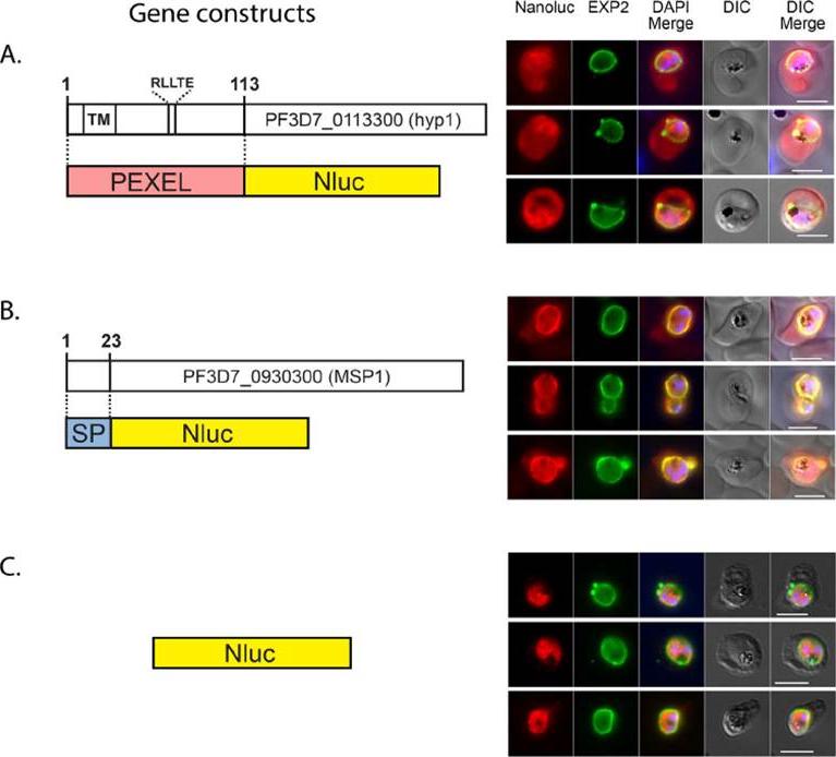
NanoLuc is targeted to the PV and to the RBC. Diagrams of gene constructs and the IFA images are shown on the left and right respectively. Nluc fused at its N-terminus to (A) the N-terminal region of an exported protein (PEXEL), (B) to a secretion signal peptide (SP) or (C) original (cytosolic). The gene encoding each fusion protein was cloned in the pEF vector. Transfected parasites were analysed by IFA using antibodies to detect Nluc and the PVM marker Exp2. DAPI was used for nuclear staining. Size bar = 5 mm.Azevedo MF, Nie CQ, Elsworth B, Charnaud SC, Sanders PR, Crabb BS, Gilson PR. Plasmodium falciparum transfected with ultra bright NanoLuc luciferase offers high sensitivity detection for the screening of growth and cellular trafficking inhibitors. PLoS One. 2014 Nov 13;9(11):e112571.
See original on MMPMore information
| PlasmoDB | PF3D7_0620400 |
| GeneDB | PF3D7_0620400 |
| Malaria Metabolic Pathways | Localisation images Pathways mapped to |
| Previous ID(s) | 2270.t00095, MAL6P1.221, PFF0995c |
| Orthologs | PBANKA_1119600 , PCHAS_1119100 , PKNH_1129800 , PVP01_1129100 , PVX_114145 , PY17X_1120700 |
| Google Scholar | Search for all mentions of this gene |