PF3D7_0604700 glyoxalase I (GILP)
Disruptability [+]
| Species | Disruptability | Reference | Submitter |
|---|---|---|---|
| P. falciparum 3D7 |
Refractory |
USF piggyBac screen (Insert. mut.) | USF PiggyBac Screen |
Mutant phenotypes [+]
None reported yet. Please press the '+' button above to add one.Imaging data (from Malaria Metabolic Pathways)
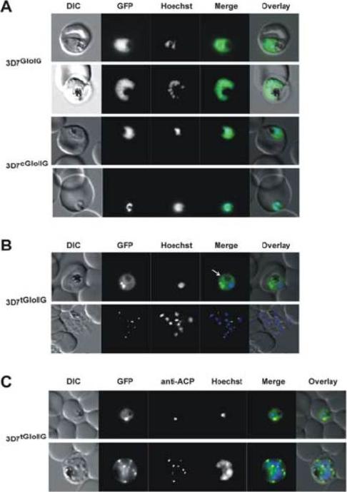
Localization of GloI, cGloII and tGloII GFP-fusion constructs in infectederythrocytes. (A) Live cell imaging of erythrocytes infected with transgenic parasite lines 3D7GloIG (upper panels) and 3D7cGloIIG (lower panels). GFP fluorescence was evident only within the body of the parasite (excluding the food vacuole). (B) Live cell imaging of erythrocytes infected with 3D7tGloIIG (apicoplast). A distinct fluorescent “dot” was seen in both trophozoite (upper panel) and merozoite (lower panel) stage parasites, indicative of an apicoplast localization. The white arrow shows additional low levels in the arasitophorous vacuole. (C) Colocalization of tGloIIGFP and the apicoplast marker ACP in fixed, immunodecorated cells. Note the high degree of overlap between tGloII-GFP and ACP.Urscher M, Przyborski JM, Imoto M, Deponte M. Distinct subcellular localization in the cytosol and apicoplast, unexpected dimerization and inhibition of Plasmodium falciparum glyoxalases. Mol Microbiol. 2010 76:92-103. Copyright John Wiley & Sons Ltd. 2010.
See original on MMPMore information
| PlasmoDB | PF3D7_0604700 |
| GeneDB | PF3D7_0604700 |
| Malaria Metabolic Pathways | Localisation images Pathways mapped to |
| Previous ID(s) | 2270.t00335, MAL6P1.50, PFF0230c |
| Orthologs | PBANKA_0103400 , PCHAS_0104100 , PKNH_1145800 , PVP01_1144200 , PVX_113400 , PY17X_0105000 |
| Google Scholar | Search for all mentions of this gene |