PF3D7_0511000 translationally-controlled tumor protein homolog (TCTP)
Disruptability [+]
| Species | Disruptability | Reference | Submitter | |
|---|---|---|---|---|
| P. falciparum 3D7 |
Possible |
USF piggyBac screen (Insert. mut.) | USF PiggyBac Screen | |
| P. berghei ANKA |
Possible |
RMgm-1507 | Imported from RMgmDB | |
| P. berghei ANKA |
Possible |
RMgm-1135 | Imported from RMgmDB | |
| P. berghei ANKA |
Possible |
PlasmoGEM (Barseq) | PlasmoGEM | |
Mutant phenotypes [+]
| Species | Stage | Phenotype | Reference | Submitter |
|---|---|---|---|---|
| P. berghei ANKA | Asexual |
Difference from wild-type |
RMgm-1507
(Most) C57BL/6 mice infected with PbNK65-hrf resolve blood stage infections. Parasites are cleared from infection around day 13/14 after infection. Clearance is dependent on the infection dose (mice infected with a low dose of 10(3)parasites did not lead to parasite clearance). In contrast, all mice infected with wild type NK65 die from severe complications (around day 20 after infection). |
Imported from RMgmDB |
| P. berghei ANKA | Asexual |
No difference |
RMgm-1135 | Imported from RMgmDB |
| P. berghei ANKA | Asexual |
Attenuated |
PlasmoGEM (Barseq) | PlasmoGEM |
| P. berghei ANKA | Gametocyte |
No difference |
RMgm-1135 | Imported from RMgmDB |
| P. berghei ANKA | Ookinete |
No difference |
RMgm-1135 | Imported from RMgmDB |
| P. berghei ANKA | Oocyst |
No difference |
RMgm-1135 | Imported from RMgmDB |
| P. berghei ANKA | Sporozoite |
No difference |
RMgm-1135 | Imported from RMgmDB |
| P. berghei ANKA | Liver |
Difference from wild-type |
RMgm-1135
Sporozoites did not display any detectable defect in gliding motility, host cell traversal or host cell invasion of HepG2 cells.HRF1 and WT developed indistinguishably inside HepG2 cells into exo-erythrocytic forms (EEF, liver stages). Therefore, HRF does not appear to be important for a basic parasite developmental step detectable in vitro.To test pre-erythrocytic stage infectivity, sporozoites were inoculated into mice by mosquito bite, intradermal or intravenous injection and emergence of blood-stage parasites monitored by FACS. Only 40 to 50% of animals became infected after injection of HRF1 sporozoites when 100% animals were infected after injection of WT parasites. Animals became patent with HRF1 blood-stage parasites on average 2 to 3 days after the WT following infection by mosquito bites or inoculation of isolated sporozoites, corresponding to a 100 to 1000-fold decrease in infectivity of mutant parasites. |
Imported from RMgmDB |
Imaging data (from Malaria Metabolic Pathways)
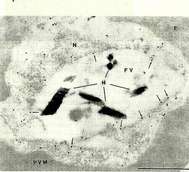
Immunoelectron microscopy of P. falciparum-infected erythrocyte using anti-TCTP. FV – food vacuole. N – nucleus. P – parasite. Bar – 1 mm. TCTP was found to be located in both the cytoplasm and the food vacuole.Bhisutthibhan J, Philbert MA, Fujioka H, Aikawa M, Meshnick SR. The Plasmodium falciparum translationally controlled tumor protein: subcellular localization and calcium binding. Eur J Cell Biol. 1999 78:665-70. Copyright Elsevier 2011.
See original on MMP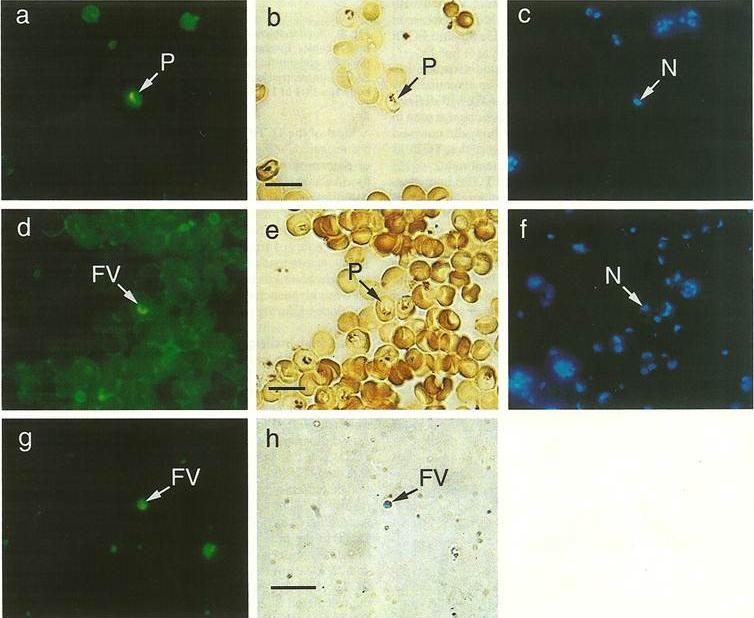
Indirect immunofluorescence of P. falcparum-infected erythrocytes using anti-TCTP antibody. Fixed culture containing trophozoites were stained with anti-TCTP Ab + FITC-labled goat anti rabbit Ab and counterstained with DAPI. a) fluorescence; b) Light microscopy; c) DAPI; d) a late trophozoite showed FITC fluorescence only in discrete vacuole; e) which can be seen to contain hemozoin; f) but to be different from nuclei; g-h) purified food vacuoles also show FITC fluorescence. FV – food vacuole. N – nucleus. P – parasite. Bar – 10 mm. TCTP was found to be located in both the cytoplasm and the food vacuole.Bhisutthibhan J, Philbert MA, Fujioka H, Aikawa M, Meshnick SR. The Plasmodium falciparum translationally controlled tumor protein: subcellular localization and calcium binding. Eur J Cell Biol. 1999 78:665-70. Copyright Elsevier 2011.
See original on MMPMore information
| PlasmoDB | PF3D7_0511000 |
| GeneDB | PF3D7_0511000 |
| Malaria Metabolic Pathways | Localisation images Pathways mapped to |
| Previous ID(s) | MAL5P1.110, PFE0545c |
| Orthologs | PBANKA_1110500 , PCHAS_1110200 , PKNH_1022600 , PVP01_1023000 , PVX_080685 , PY17X_1111700 |
| Google Scholar | Search for all mentions of this gene |