PF3D7_0501100 heat shock protein 40, type II (HSP40)
Disruptability [+]
| Species | Disruptability | Reference | Submitter |
|---|---|---|---|
| P. falciparum 3D7 |
Possible |
18614010 | Theo Sanderson, Wellcome Trust Sanger Institute |
| P. falciparum 3D7 |
Refractory |
USF piggyBac screen (Insert. mut.) | USF PiggyBac Screen |
Mutant phenotypes [+]
None reported yet. Please press the '+' button above to add one.Imaging data (from Malaria Metabolic Pathways)
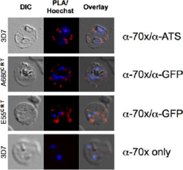
PfHsp70-x interacts with exported Hsp40s and PfEMP1. A. proximity ligation assay (PLA) of (upper panel) PfHsp70-x/PfEMP1, (second panel) PfHsp70-x/A660-GFP (PFA0660w), (third panel) PfHsp70-x/E55-GFP (PFE0055c). A red signal indicates a positive interaction. Lower panel shows negative control. PfHsp70-x appears to closely interact with the ATS of PfEMP1, and both exported Hsp40s studied.Külzer S, Charnaud S, Dagan T, Riedel J, Mandal P, Pesce ER, Blatch GL, Crabb BS, Gilson PR, Przyborski JM. Plasmodium falciparum-encoded exported hsp70/hsp40 chaperone/co-chaperone complexes within the host erythrocyte. Cell Microbiol. 2012 14(11):1784-95
See original on MMP
Parasites were tagged by single cross-over homologous recombination, the endogenous gene locus to include the GFP coding sequence. PFE55INT. Co-immunofluorescence analysis on erythrocytes infected with PFE55INT, using anti-sera directed against STEVOR (A), MAHRP1 MAL13P1.413 (B), KAHRP PFB0100c (C). Fluorescence channels are shown individually in black/white for highest contrast. All images are maximal projections of Z-stack serial sections. In merge image green, GFP; red, STEVOR (A), MAHRP (B), KAHRP (C) or ATS domain of PfEMP1 (D); blue, Hoechst. Inset shows enlargement of merge (white box). The fusion protein labelled punctate structures within the infected erythrocytebut clearly not in Maurer’s clefts.Külzer S, Rug M, Brinkmann K, Cannon P, Cowman A, Lingelbach K, Blatch GL, Maier AG, Przyborski JM. Parasite encoded Hsp40 proteins define novel mobile structures in the cytosol of the P. falciparum infected erythrocyte. Cell Microbiol. 2010 12(10):1398-420 Copyright John Wiley & Sons Ltd. 2010.
See original on MMP
(A) Live cell imaging of co-transfectants expressing saGFP (self-assembling split GFP) fragments in various cellular compartments. In merge and overlay: green, GFP; blue, Hoechst. (B) Vectors available for analysis of cellular compartmentalisation using saGFP. Numbers in targeting sequence refer to N-terminal amino acids. B, Blasticidin-S-deaminase resistance cassette; D, hDHFR resistance cassette. *Can be used to generate fusions as multiple cloning sites situated in front of saGFP coding sequence. Külzer S, Petersen W, Baser A, Mandel K, Przyborski JM. Use of self-assembling GFP to determine protein topology and compartmentalisation in the Plasmodium falciparum-infected erythrocyte. Mol Biochem Parasitol. 2012 187(2):87-90.
See original on MMP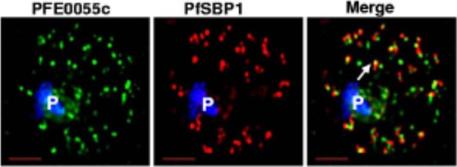
Single optical section of a trophozoite-infected erythrocyte fixed and probed with peptide antibodies to PFE0055c (green) and the cleft protein SBP1 (red). Arrow in merge image shows proximal location of PFE0055c to clefts.Bhattacharjee S, van Ooij C, Balu B, Adams JH, Haldar K. Maurer's clefts of Plasmodium falciparum are secretory organelles that concentrate virulence protein reporters for delivery to the host erythrocyte. Blood. 2008 111:2418-26.
See original on MMP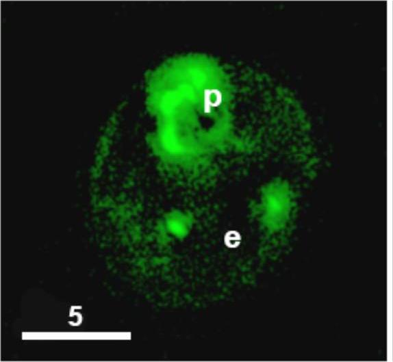
Export of a secretome-predicted parasite HSP40-GFP chimera to the erythrocyte. Projection (0o) of a live cell expressing PfHSP40GFP. Green, GFP; p, parasite; e, erythrocyte. Scale bar 5μm.Hiller NL, Akompong T, Morrow JS, Holder AA, Haldar K. Identification of a stomatin orthologue in vacuoles induced in human erythrocytes by malaria parasites. A role for microbial raft proteins in apicomplexan vacuole biogenesis. J Biol Chem. 2003 278:48413-21.
See original on MMP
Single optical section of a trophozoite-infected erythrocyte fixed and probed with peptide antibodies to PFE0055c (green) and the cleft protein SBP1 (red). Arrow in merge image shows proximal location of PFE0055c to clefts.Bhattacharjee S, van Ooij C, Balu B, Adams JH, Haldar K. Maurer's clefts of Plasmodium falciparum are secretory organelles that concentrate virulence protein reporters for delivery to the host erythrocyte. Blood. 2008 111:2418-26.
See original on MMP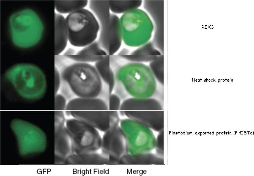
GFP fusions to three genes (PFI1780w, PFE0055c, PFI1755c) whose products are exported into the red blood cell cytosol.Sargeant TJ, Marti M, Caler E, Carlton JM, Simpson K, Speed TP, Cowman AF. Lineage-specific expansion of proteins exported to erythrocytes in malaria parasites. Genome Biol. 2006;7(2):R12;
See original on MMP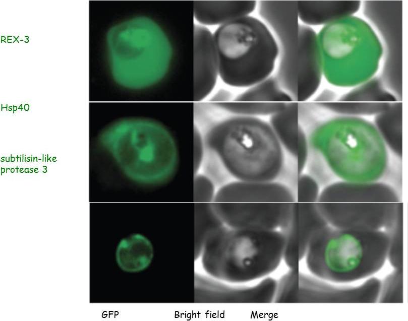
Parasites were transfected with the GFP chimeras of the 3 genes. Experimental verification of their sub-cellular localization indicated that REX-3 and Hsp40 were exported into the host cell cytosol while the protease accumulated in the parasitophorous vacuole.Sargeant TJ, Marti M, Caler E, Carlton JM, Simpson K, Speed TP, Cowman AF. Lineage-specific expansion of proteins exported to erythrocytes in malaria parasites. Genome Biol. 2006; 7(2):R12.
See original on MMPMore information
| PlasmoDB | PF3D7_0501100 |
| GeneDB | PF3D7_0501100 |
| Malaria Metabolic Pathways | Localisation images Pathways mapped to |
| Previous ID(s) | MAL5P1.12, PF3D7_0501100.1, PF3D7_0501100.2, PFE0055c |
| Orthologs | |
| Google Scholar | Search for all mentions of this gene |