PF3D7_0500800 mature parasite-infected erythrocyte surface antigen (MESA)
Disruptability [+]
| Species | Disruptability | Reference | Submitter |
|---|---|---|---|
| P. falciparum 3D7 |
Possible |
USF piggyBac screen (Insert. mut.) | USF PiggyBac Screen |
Mutant phenotypes [+]
None reported yet. Please press the '+' button above to add one.Imaging data (from Malaria Metabolic Pathways)
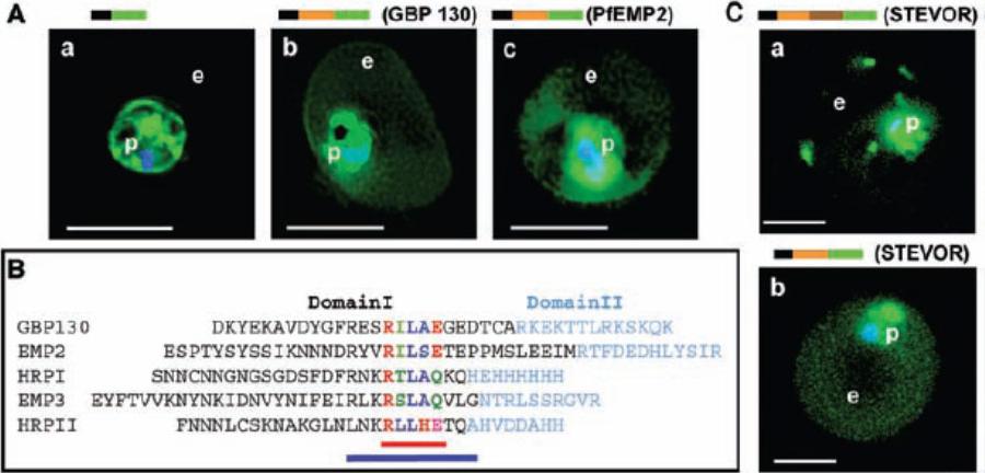
Identification of a conserved motif of 11 amino acids in VTSs of parasite proteins exported to the erythrocyte. (A) VTS of proteins PfGBP 130 and PfEMP2 target GFP to the erythrocyte. Projections (0-) of live cells expressing GFP chimeras of a SS alone (a), SSVTSGBP130 (b), and SSVTSEMP2 (c). (B) Alignment of MEME motifs in VTSs of indicated five exported soluble proteins. Red bar underlines 5–amino acid MEME motif 1. Blue bar underlines 11–amino acid MEME motif 2. (C) Projections (0-) of live cells expressing GFP chimeras of full-length STEVOR (a) and SSVTSSTEVOR (b).Hiller NL, Bhattacharjee S, van Ooij C, Liolios K, Harrison T, Lopez-Estraño C, Haldar K. A host-targeting signal in virulence proteins reveals a secretome in malarial infection. Science. 2004 306(5703):1934-7.
See original on MMP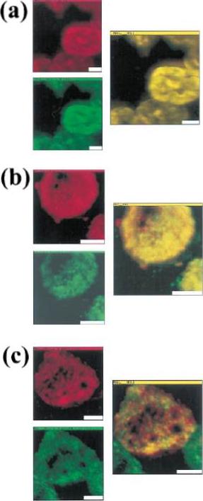
Colocalization of malarial and host proteins in the erythrocyte membrane. A blood smear of erythrocytes infected with the trophozoite stage parasites was fixed and then reacted with two different antibodies (from rabbit and from mouse) against the proteins under study. Secondary antibodies labeled with FITC (anti-rabbit) and TR (anti-mouse) were used, respectively. The left column shows the two simultaneously acquired fluorescence channels by NSOM (near-field scanning optical microscope) dualcolor imaging. The center column presents the corresponding overlay of the fluorescence images (a) Control experiment: primary antibodies against KAHRP. (b) Colocalization of MESA and protein 4.1 (a host cell membrane protein). (c) Colocalization of KAHRP and protein 4.1. Scale bars 5 2 mm.Enderle T, Ha T, Ogletree DF, Chemla DS, Magowan C, Weiss S. Membrane specific mapping and colocalization of malarial and host skeletal proteins in the Plasmodium falciparum infected erythrocyte by dual-color near-field scanning optical microscopy. Proc Natl Acad Sci U S A. 1997 94:520-5.
See original on MMP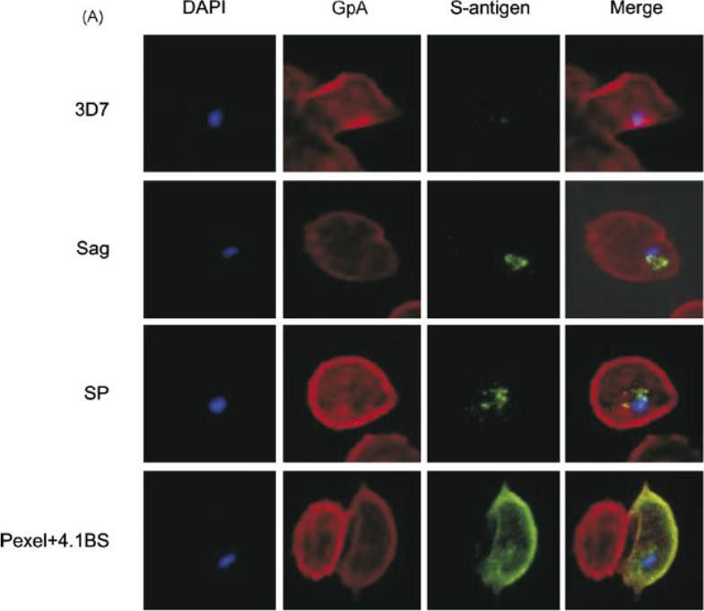
A mouse antibody to RBC membrane protein Glycophorin A (GpA) was used as a positive control for the IFA experiments. The images show that the proteins expressed by control line (3D7 Sag) which contains S-antigen alone (with its endogenous signal peptide) remained confined within the parasite or the PV, and was not exported into the RBC cytosol. The protein expressed by 3D7 SP (expressing exon 1 of MESA fused to S-antigen) was also retained within the PV, indicating that the embedded signal peptide of MESA is not sufficient to transport the protein across the PVM. The image for 3D7 Pexel+4.1BS, which contains the putative Pexel and the 4.1R-binding site, shows that the protein was trafficked outside the parasite and appeared to co-localise with GpA at the RBC membrane. Transfectant parasite line 3D7 D4.1BS (lacking the 4.1R-binding site) showed a vastly different staining pattern to that of 3D7 Pexel+4.1BS. It appeared that the D4.1BS/S-antigen chimera was being transported outside the parasite, indicating that the Pexel is functional and is the minimum sequence required for passage through the PVM into the RBC cytosol.Black CG, Proellocks NI, Kats LM, Cooke BM, Mohandas N, Coppel RL. In vivo studies support the role of trafficking and cytoskeletal-binding motifs in the interaction of MESA with the membrane skeleton of Plasmodium falciparum-infected red blood cells. Mol Biochem Parasitol. 2008 160:143-7. Copyright Elsevier 2009.
See original on MMP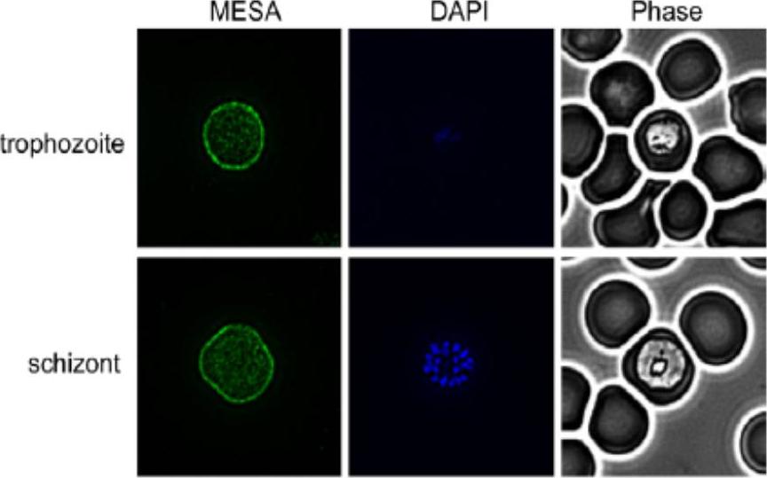
Indirect Immunofluorescence Assay (IFA) was performed using anti-MESA antiserum. Representative IFAs are shown. More diffuse RBC membrane labeling of MESA was detected on trophozoites and schizonts. No labeling was detected in rings (data not shown). PfEMP2/MESA localizes to the RBC membrane.Waller KL, McBride SM, Kim K, McDonald TV. Characterization of two putative potassium channels in Plasmodium falciparum. Malar J. 2008 Jan 24;7:19.
See original on MMP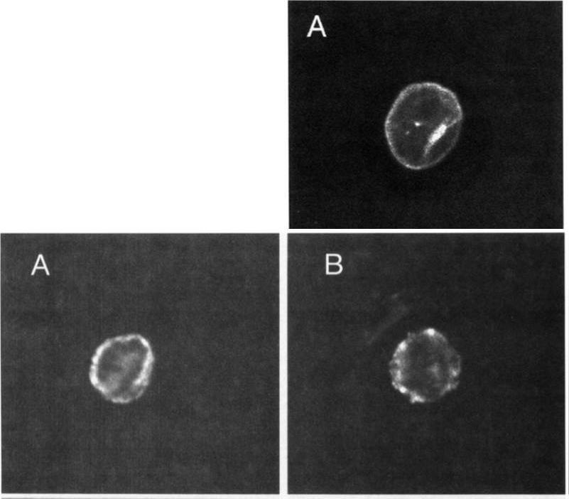
Upper: Localization of MESA. Thin blood smears of mature trophozoite-infected erythrocytes were made from in vitro cultures, air-dried, and fixed with acetonelrnethanol. They were reacted at room temperature for 45 minutes with anti-MESA diluted 1:lOO in PES, washed 3 times in PES, and reacted for 45 minutes with fluorescein isothiocyanate-conjugated goat-antirabbit IgG diluted 1:lOO in PBS. Slides were again washed 3 times in PBS and mounted with SlowFade and a coverslip for examination with a confocal laser scanning microscope equipped with Nikon epifluorescence. Lower: Localization of KAHRP. Cells were prepared and examined as above except that in this case they were reacted with MoAb 89 (antiknob-associated histidine-rich protein) diluted 120 in PBS, followed by fluorescein isothiocyanate-conjugated goat-antimouse IgG diluted 1:lOO in PBS.Magowan C, Coppel RL, Lau AO, Moronne MM, Tchernia G, Mohandas N. Role of the Plasmodium falciparum mature-parasite-infected erythrocyte surface antigen (MESA/PfEMP-2) in malarial infection of erythrocytes.Blood. 1995 86:3196-204.
See original on MMP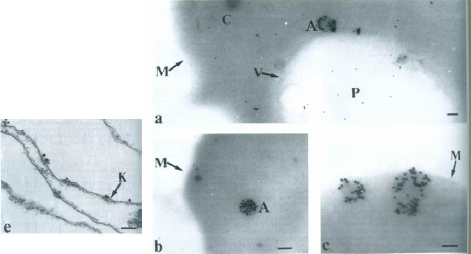
Right: Subcellular localization of PfEMP2 using IgM mAb 4H9.1 by cryo-thin-section immunoelectron microscopy. Electron-dense clumps are seen associated with PfEMP2 antigen,. Bar 0.5 mm. P – parasite; C – erythrocyte cytoplasm; V – parasitophophorous vacuole membrane; M – infected erythrocyte surface membrane.e. Sub-membrane area of knob (K) identified by concavity of surface membrane and electron-dense material. Bar 0.25 mm.Howard RJ, Lyon JA, Uni S, Saul AJ, Aley SB, Klotz F, Panton LJ, Sherwood JA, Transport of an Mr approximately 300,000 Plasmodium falciparum protein (Pf EMP 2) from the intraerythrocytic asexual parasite to the cytoplasmic face of the host cell membrane. J Cell Biol. 1987 May;104(5):1269-80.
See original on MMP
PfGECO is external to Pfs16 and internal to MESA. Co-localization IFA was performed on gametocyte stages I-IV using anti-PfGECO serum (red) and either (A) anti-Pfs16 (green) serum or (B) anti-MESA (green) serum. Also shown is the parasite nucleus stained with DAPI (blue), the bright field (BF) and the merged images.Morahan BJ, Strobel C, Hasan U, Czesny B, Mantel PY, Marti M, Eksi S, Williamson KC. Functional analysis of the exported type IV HSP40 protein PfGECO in P. falciparum gametocytes. Eukaryot Cell. 2011 10(11):1492-503.
See original on MMP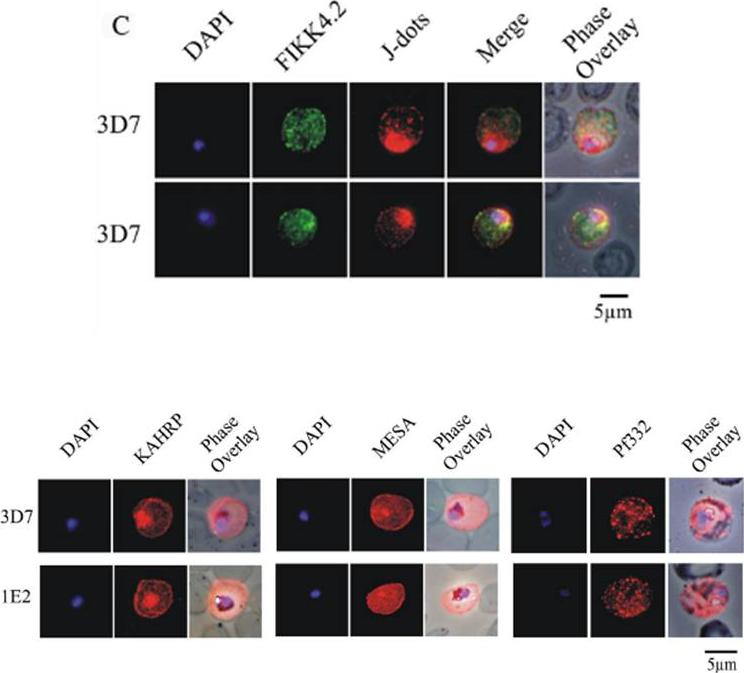
(C) Immunofluorescence analysis of red blood cells infected with parental 3D7 parasites co-labelled with a-FIKK4.2 and a-PFB0090c (J-dots) antibodies. Merge and phase overlay images indicate that FIKK4.2 and J-dots do not co-localise in infected red blood cells. In all panels, parasite nuclei are counterstained with DAPI. (D) Immunofluorescence analysis of red blood cells infected with either parental 3D7 or FIKK4.2 knockout (1E2) parasites. Infected red blood cells were incubated with antibodies raised against the knob-associated histidine-rich protein (KAHRP), mature-parasite-infected erythrocyte surface antigen (MESA) or P. falciparum antigen 332 (Pf332). Parasite nuclei were counterstained with DAPI. KAHRP, MESA and Pf332 as three examples of other very well described P. falciparum-encoded exported proteins.Kats LM, Fernandez KM, Glenister FK, Herrmann S, Buckingham DW, Siddiqui G, Sharma L, Bamert R, Lucet I, Guillotte M, Mercereau-Puijalon O, Cooke BM. An exported kinase (FIKK4.2) that mediates virulence-associated changes in Plasmodium falciparum-infected red blood cells. Int J Parasitol. 2014 Feb 14. [Epub ahead of print]
See original on MMP
Localization of LyMP in iRBCs. A) Localization of LyMP in trophozoites, with either anti-LyMP (green) or anti-HA (red) showing that LyMP is exported into the RBC cytosol. 4’,6’-Diamidino-2-phenylidole (DAPI) is a nuclear stain (blue). B) Colocalization of LyMP (green) with a variety of iRBC markers (red) that are indicated in the panel. DAPI is a nuclear stain (blue). C) High-resolution structured illumination fluorescence microscopy (OMX) with anti-LyMP (green) and either anti-KAHRP, anti-MESA, or anti-spectrin (red) colocalizations. Corresponding overlay images are shown for all. D) Immunoelectron micrograph of gold-labeled Ha-tagged parasites. Red asterisks indicate knobs and the arrow indicates gold labeling of LyMP at the RBC membrane skeleton. P, parasite; MC, Maurer’s clefts. LyMP showed a pattern of distribution similar to that for MESA, KAHRP. LyMP did not consistently colocalize with MCs or to other punctate structures previously identified in the iRBC cytosol such as those seen for FIKK4.2 (R45) or for J-dots (KAHsp40).Proellocks NI, Herrmann S, Buckingham DW, Hanssen E, Hodges EK, Elsworth B, Morahan BJ, Coppel RL, Cooke BM. A lysine-rich membrane-associated PHISTb protein involved in alteration of the cytoadhesive properties of Plasmodium falciparum-infected red blood cells. FASEB J. 2014 Apr 4. [Epub ahead of print]
See original on MMPMore information
| PlasmoDB | PF3D7_0500800 |
| GeneDB | PF3D7_0500800 |
| Malaria Metabolic Pathways | Localisation images Pathways mapped to |
| Previous ID(s) | MAL5P1.9, PFE0040c |
| Orthologs | |
| Google Scholar | Search for all mentions of this gene |