PF3D7_0424400 surface-associated interspersed protein 4.2 (SURFIN 4.2) (SURF4.2)
Disruptability [+]
| Species | Disruptability | Reference | Submitter |
|---|---|---|---|
| P. falciparum 3D7 |
Possible |
18614010 host erythrocyte |
Theo Sanderson, Wellcome Trust Sanger Institute |
| P. falciparum 3D7 |
Possible |
USF piggyBac screen (Insert. mut.) | USF PiggyBac Screen |
Mutant phenotypes [+]
None reported yet. Please press the '+' button above to add one.Imaging data (from Malaria Metabolic Pathways)
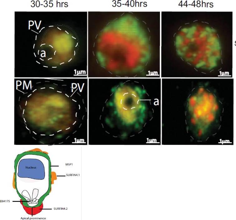
Localization of SURFIN4.1 by immunofluorescence staining on air-dried monolayers from 3D7S8 parasite strain. Air dried monolayers probed with rabbit anti-SURFIN4.1 on 3D7S8 pRBC. Propidium iodide (red) was used to stain the parasite nucleus and SURFIN4.1 and SURFIN4.2 (which is encoded by the same gene) proteins were stained green using anti-rabbit Alexa 488. SURFIN4.1 localizes within the parasitophorous vacuole (PV) and is observed from approximately 30 hrs post invasion. SURFIN4.1 was observed as a green dot above the food vacuole (a), at 30–35 hrs parasite stages. The protein was spread around the parasitophorous vacuole (PV) at 35–40 hrs parasite stages and in the mature schizont (44–48 hrs) SURFIN4.1 was observed between the dividing merozoites. During the trophozoite and early schizont stages SURFIN4.2 shows a similar pattern of staining as SURFIN4.1.Mphande FA, Ribacke U, Kaneko O, Kironde F, Winter G, Wahlgren M. SURFIN4.1, a schizont-merozoite associated protein in the SURFIN family of Plasmodium falciparum. Malar J. 2008 Jul 1;7:116. PMID:
See original on MMP
SURFIN accumulates within the parasitophorous vacuole in late schizonts and is associated with released merozoites. (A) Alexa 488–labeled SURFIN4.2 (green) detected with anti-S1.3 antibodies in parasites developing from late trophozoite stage to schizonts. PI staining (red) was used to visualize nuclei. (B–D) SURFIN4.2 was found localized in MAM and the PM of late and bursting schizonts (48 h), and associated with the released merozoite surface. Dashed circles indicate the area of the IE. (B) IFA with anti-S1.3 + PI on bursting schizonts. IFA (anti-rSURFIN4.2) and immunoelectron microscopy (anti-S1.4 Ig) on late schizonts (C) and released merozoites (D). SURFIN associated with PM or merozoite-associated material is indicated by arrows. (E) SURFIN4.2 did not colocalize with the microneme AMA-1. A detailed analysis of SURFIN4.2 (anti-S1.3 antibodies) and AMA-1 in relation to the nuclei and to each other in merozoites is shown. (B and E) A schematic representation of the antigen localization is shown on the right. Bars: IFA, 2 mm; EM, 0.5 mm.Winter G, Kawai S, Haeggström M, Kaneko O, von Euler A, Kawazu S, Palm D, Fernandez V, Wahlgren M. SURFIN is a polymorphic antigen expressed on Plasmodium falciparum merozoites and infected erythrocytes. J Exp Med. 2005 201:1853-63.
See original on MMP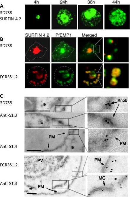
SURFINs expressed in 3D7S8 and FCR3S1.2 are cotransported with PfEMP1 to the host PM. (A) Indirect immunofluorescence assay (IFA) on air-dried monolayers of 3D7S8 IEs at various stages of maturation with SURFIN4.2-directed anti-S1.3 antibodies. Antibodies detected SURFIN4.2 in 25% of examined parasites at 24 h in dot-like transport vesicles outside the parasitophorous vacuole. (B) Dual localization of TRITC-labeled SURFIN4.2 (red) and Alexa 488–labeled PfEMP1 (green) showed that SURFIN4.2 and PfEMP1 are colocalized in cytosolic structures. An expressed SURFIN was detected in 90% of trophozoite-stage FCR3S1.2 IEs and colocalized with PfEMP1 in large multimeric vesicles. Dashed circles indicate the area of the IE. (C) Immunoelectron microscopy studies on 3D7S8 and FCR3S1.2 IEs. SURFIN detected with either anti-S1.3 serum or affinity purified anti-S1.4 immunoglobulins is present in the PM, as well as in the erythrocyte cytosol associated with Maurer’s clefts (MC) as indicated by arrows. Bars: (A and B), 2 mm; (C), 0.5 mm.Winter G, Kawai S, Haeggström M, Kaneko O, von Euler A, Kawazu S, Palm D, Fernandez V, Wahlgren M. SURFIN is a polymorphic antigen expressed on Plasmodium falciparum merozoites and infected erythrocytes. J Exp Med. 2005 201:1853-63.
See original on MMP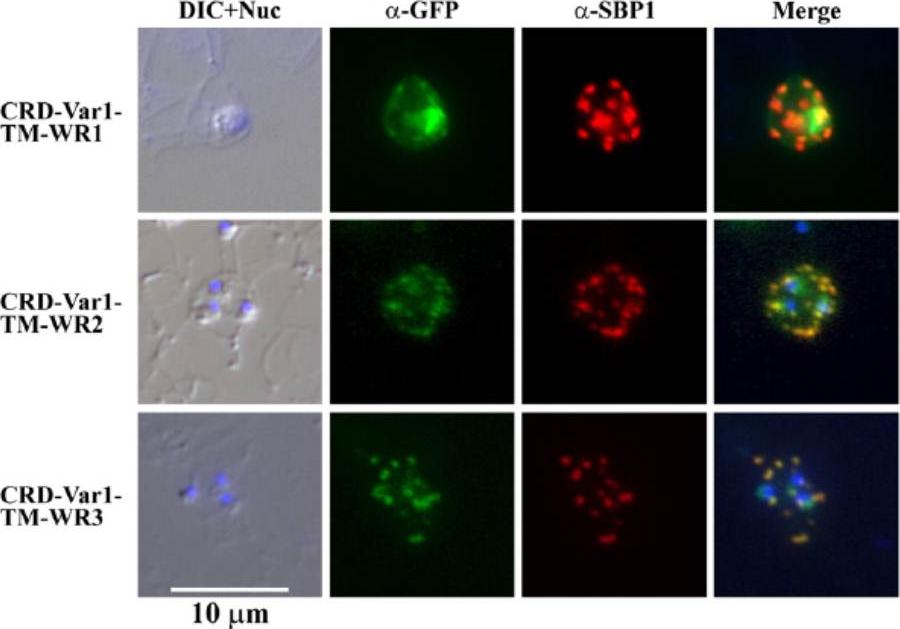
Indirect immunofluorescence assay of three mini-SURFIN4.2 proteins. Double staining IFA for 3 mini-SURFIN4.2-expressing transfectants is shown. α-GFP and α-SBP1 indicate anti-GFP (mini-SURFIN4.2) and anti-SBP1 (Maurer's cleft protein). Negative controls using normal rabbit antibody did not produce detectable signals (not shown). CRD, cysteine-rich domain; TM, transmembrane; and Var1, variable region 1; WR, tryptophan-rich.Alexandre JS, Yahata K, Kawai S, Torii M, Kaneko O. PEXEL-independent trafficking of Plasmodium falciparum SURFIN(4.2) to the parasite-infected red blood cell and Maurer's clefts. Parasitol Int. 2011 60(3):313-20.
See original on MMP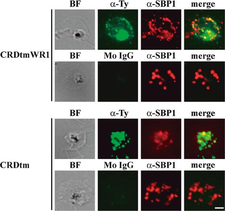
Indirect immunofluorescence assay of recombinant CRDtmWR1 (recombinant SURFIN4.2 proteins containing intracellular region) and CRDtm (recombinant SURFIN4.2 proteins not containing intracellular region). CRDtmWR1 and CRDtm were dual stained with mouse anti-Ty antibody (α-Ty, green; Monoclonal antibody raised in mouse against the Ty1 tag (amino acid sequence EVHTNQDPLD) for recombinant SURFIN4.2 and rabbit anti-PfSBP1 serum (α-SBP1, red) for Maurer’s clefts. Dual staining negative control images with mouse normal IgG (Mo IgG) and anti-PfSBP1 are also shown. Bright field images (BF) are shown, as well as merged images (merge) of α-Ty, α-SBP1, and DAPI (blue) DNA staining. The bar = 2 μm.Kagaya W, Miyazaki S, Yahata K, Ohta N, Kaneko O. The Cytoplasmic Region of Plasmodium falciparum SURFIN4.2 Is Required for Transport from Maurer's Clefts to the Red Blood Cell Surface. Trop Med Health. 2015 Dec;43(4):265-72.
See original on MMPMore information
| PlasmoDB | PF3D7_0424400 |
| GeneDB | PF3D7_0424400 |
| Malaria Metabolic Pathways | Localisation images Pathways mapped to |
| Previous ID(s) | 3D7Surf4.2, MAL4P1.227, PFD1160w |
| Orthologs | |
| Google Scholar | Search for all mentions of this gene |