PF3D7_0424200 reticulocyte binding protein homologue 4 (RH4)
Disruptability [+]
| Species | Disruptability | Reference | Submitter |
|---|---|---|---|
| P. falciparum 3D7 |
Possible |
16123303 | Theo Sanderson, Wellcome Trust Sanger Institute |
| P. falciparum 3D7 |
Refractory |
USF piggyBac screen (Insert. mut.) | USF PiggyBac Screen |
Mutant phenotypes [+]
None reported yet. Please press the '+' button above to add one.Imaging data (from Malaria Metabolic Pathways)
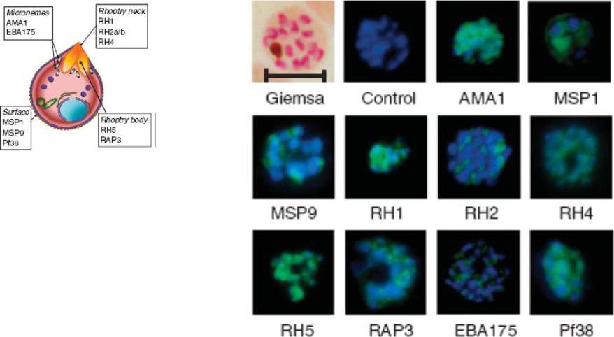
Indirect immunofluorescence images of P. falciparum schizonts stained with rabbit IgG (green) induced by 10 viral-vectored vaccines expressing malaria antigens, and negative control vectors lacking a malaria antigen. Nuclei are counterstained with 4,6-diamidino-2-phenylindole (blue). All sera were tested against 3D7 strain parasites, with the exception of anti-PfRH1 for which FVO strain parasites were used. All images to same scale as Giemsa stained image (top left, on which scale bar indicates 5 μm).Douglas AD, Williams AR, Illingworth JJ, Kamuyu G, Biswas S, Goodman AL, Wyllie DH, Crosnier C, Miura K, Wright GJ, Long CA, Osier FH, Marsh K, Turner AV, Hill AV, Draper SJ. The blood-stage malaria antigen PfRH5 is susceptible to vaccine-inducible cross-strain neutralizing antibody. Nat Commun. 2011 2:601.
See original on MMP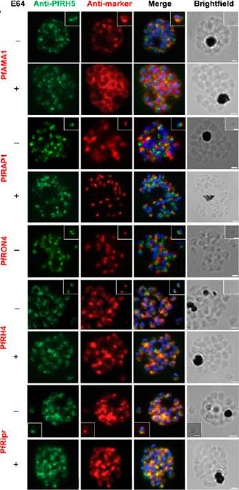
Localization of PfRH5 was assessed by indirect IFA using anti-PfRH5 polyclonal rabbit serum (green). Fixed and permeabilized schizonts with (+) or without (2) E64 treatment or free merozoites (inset) of 3D7 clone P. falciparum were costained with mouse Abs (red) to mark various organelles: PfAMA1 polyclonal (microneme), PfRAP1 mAb (rhoptry body), or PfRON4 mAb (rhoptry neck); or polyclonal mouse serum against further Ags: PfRH4 and PfRipr. Figures show the merge of the dual staining Abs and nuclei stained with DAPI (blue), as well as the brightfield view. Scale bars, 1 mm. anti-PfRH5 rabbit Abs (8) do not colocalize with conventional markers of the rhoptry bulb, rhoptry neck, or micronemes (PfRAP1, PfRON4, and PfAMA1, respectively) in permeabilized schizonts or free merozoites of 3D7 clone P. falciparum parasites. Minimal colocalization in schizonts with PfRH2a/b (data not shown) and also with PfRH4, described as a marker of the rhoptry tip, but significant colocalization with PfRipr in merozoites and late-stage schizonts, especially following treatment with the cysteine protease inhibitor E64, which prevents merozoite release by inhibiting schizont rupture. Impossible to detect PfRH5 on the merozoite surface in any assay, with staining only successful following permeabilization .Douglas AD, Williams AR, Knuepfer E, Illingworth JJ, Furze JM, Crosnier C, Choudhary P, Bustamante LY, Zakutansky SE, Awuah DK, Alanine DG, Theron M, Worth A, Shimkets R, Rayner JC, Holder AA, Wright GJ, Draper SJ. Neutralization of Plasmodium falciparum Merozoites by Antibodies against PfRH5. J Immunol. 2014 192(1):245-58.
See original on MMP
Subnuclear Localization of PfRh4 Varies with Invasion Phenotype Switching. (A) Representative images of DNA FISH measuring the colocalization of active and silent PfRh loci (green) with Rep20 (red). (D) Schematic and representative images of DNA FISH localizing PfRh4 (green) and a plasmid containing transcriptionally active hDHFR (red) and localized to the nuclear periphery with Rep20 (gray).Coleman BI, Ribacke U, Manary M, Bei AK, Winzeler EA, Wirth DF, Duraisingh MT. Nuclear Repositioning Precedes Promoter Accessibility and Is Linked to the Switching Frequency of a Plasmodium falciparum Invasion Gene. Cell Host Microbe. 2012 12(6):739-50. PMID:
See original on MMP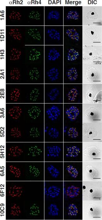
Localization of native PfRh4 was assessed by wide-field immunofluorescence assay using the anti-PfRh4 mAbs. PfRh4 (green) was co-stained with rhoptry protein, PfRh2 (red) and nuclei-stain DAPI (blue). DIC shows the differential interference contrast view of the same field. Scale bar = 5 μm. PfRh4, together with the other PfRh family of proteins, localize to the rhoptries of P. falciparum parasites in an apical localization that predominantly co-localized with PfRh2 another rhoptry protein.Lim NT, Harder MJ, Kennedy AT, Lin CS, Weir C, Cowman AF, Call MJ, Schmidt CQ, Tham WH. Characterization of Inhibitors and Monoclonal Antibodies that Modulate the Interaction between Plasmodium falciparum Adhesin PfRh4 with its Erythrocyte Receptor Complement Receptor 1. J Biol Chem. 2015 Aug 31. [Epub ahead of print]
See original on MMP
Specific amino acid residues in the cytoplasmic domain are essential for PfRh4 function. Localization of PfRh4 and PfRh2 in Rh4-WT tail, RH4-AMA1 tail (PfRh4 tail was replaced with the cytoplasmic sequence of AMA1), Rh4-mut4tail ) contains the mutations S1667A, S1674A, Y1680A and Y1684A) and RH4-mut5tail (contains similar mutations with an addition mutation at S1652A within the PfRh4 cytoplasmic tail) lines as detected using anti-PfRh4 monoclonal and anti-PfRh2 polyclonal antibodies. Parasite nuclei were stained with DAPI. Rh4-mut4 tail and Rh4-mut5 tail expressed PfRh4 and all showed an apical localization similar to rhoptry protein PfRh2Tham WH, Lim NT, Weiss GE, Lopaticki S, Ansell BR, Bird M, Lucet I, Dorin-Semblat D, Doerig C, Gilson PR, Crabb BS, Cowman AF. Plasmodium falciparum Adhesins Play an Essential Role in Signalling and Activation of Invasion into Human Erythrocytes. PLoS Pathog. 2015 Dec 22;11(12):e1005343.
See original on MMPMore information
| PlasmoDB | PF3D7_0424200 |
| GeneDB | PF3D7_0424200 |
| Malaria Metabolic Pathways | Localisation images Pathways mapped to |
| Previous ID(s) | MAL4P1.225, PFD1150c |
| Orthologs | |
| Google Scholar | Search for all mentions of this gene |