PF3D7_0424100 reticulocyte binding protein homologue 5 (RH5)
Disruptability [+]
| Species | Disruptability | Reference | Submitter |
|---|---|---|---|
| P. falciparum 3D7 |
Refractory |
19000690 | Theo Sanderson, Wellcome Trust Sanger Institute |
| P. falciparum 3D7 |
Refractory |
USF piggyBac screen (Insert. mut.) | USF PiggyBac Screen |
Mutant phenotypes [+]
| Species | Stage | Phenotype | Reference | Submitter |
|---|---|---|---|---|
| P. falciparum 3D7 | Asexual |
Invasion defect |
36396942 (Conditional)
Analysis of parasite growth showed they were not able to expand, indicating the function of each protein was essential (Fig. 1d–f and Extended Data Fig. 1g–i) and that the schizont to ring stage transition was blocked, consistent with these proteins being required for invasion (Fig. (Fig.2a2a). |
Theo Sanderson, Francis Crick Institute |
Imaging data (from Malaria Metabolic Pathways)
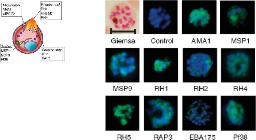
Indirect immunofluorescence images of P. falciparum schizonts stained with rabbit IgG (green) induced by 10 viral-vectored vaccines expressing malaria antigens, and negative control vectors lacking a malaria antigen. Nuclei are counterstained with 4,6-diamidino-2-phenylindole (blue). All sera were tested against 3D7 strain parasites, with the exception of anti-PfRH1 for which FVO strain parasites were used. All images to same scale as Giemsa stained image (top left, on which scale bar indicates 5 μm).Douglas AD, Williams AR, Illingworth JJ, Kamuyu G, Biswas S, Goodman AL, Wyllie DH, Crosnier C, Miura K, Wright GJ, Long CA, Osier FH, Marsh K, Turner AV, Hill AV, Draper SJ. The blood-stage malaria antigen PfRH5 is susceptible to vaccine-inducible cross-strain neutralizing antibody. Nat Commun. 2011 2:601.
See original on MMP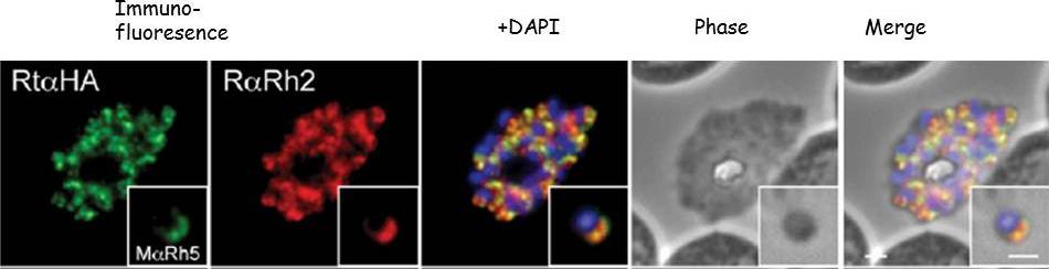
Immunofluorescence and phase contrast images of late schizonts or free merozoites (insets) to colocalise PfRh5 with PfRh2a/b. Each panel from left top right corresponds to anti-HA antibodies (to detect PhRh5), rabbit anti-PfRh2a/b, overlay of each with DAPI nuclear stain, phase contrast and overlay of all images. Insets show individual merozoites. Scale bars = 1 mM. Baum J, Chen L, Healer J, Lopaticki S, Boyle M, Triglia T, Ehlgen F, Ralph SA, Beeson JG, Cowman AF. Reticulocyte-binding protein homologue 5 - An essential adhesin involved in invasion of human erythrocytes by Plasmodium falciparum. Int J Parasitol. 2008 39(3):371-80 Copyright Elsevier 2009
See original on MMP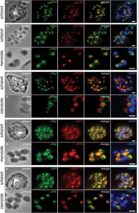
PfRipr localizes to the apical end of merozoites. a) Rabbit polyclonal anti-PfRip antibody (anti-PfRipr/1) recognizes HA-tagged PfRiprHA. b) PfRipr does not co-localize with the rhoptry neck protein RON4 in schizonts. c) PfRipr does not co-localize with the rhoptry neck protein RON4 in merozoites. d) PfRipr does not co-localize with the rhoptry bulb protein, RAP1 in schizonts. e) PfRipr does not co-localize with the rhoptry bulb protein, RAP1 in merozoites. f) PfRipr partially co-localizes with PfRh5 in the schizonts. g) PfRipr mainly co-localizes with PfRh5 in purified merozoites. h) PfRipr co-localizes with the micronemal marker, EBA175, in schizonts. i) PfRipr does not co-localize with the micronemal marker, EBA175, in merozoites.Chen L, Lopaticki S, Riglar DT, Dekiwadia C, Uboldi AD, Tham WH, O'Neill MT, Richard D, Baum J, Ralph SA, Cowman AF. An EGF-like protein forms a complex with PfRh5 and is required for invasion of human erythrocytes by Plasmodium falciparum. PLoS Pathog. 2011 7(9):e1002199.
See original on MMP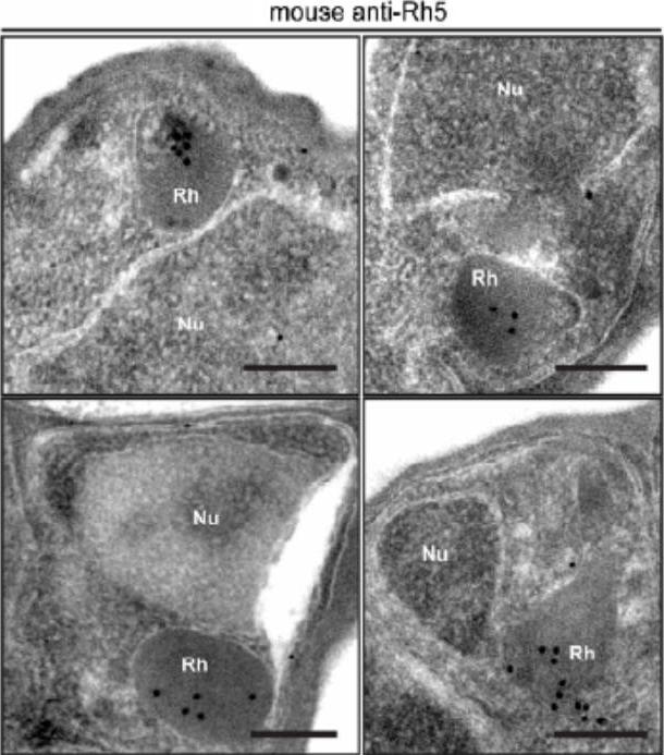
Immuno-electron microscopy of late schizonts localising PfRh5 (using both anti-PfRh5 and anti-HA in the tagged line) to the merozoite rhoptries. Scale bars = 0.2 mm.Baum J, Chen L, Healer J, Lopaticki S, Boyle M, Triglia T, Ehlgen F, Ralph SA, Beeson JG, Cowman AF. Reticulocyte-binding protein homologue 5 - An essential adhesin involved in invasion of human erythrocytes by Plasmodium falciparum. Int J Parasitol. 2009 39:371-80. Copyright Elsevier.
See original on MMP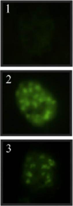
Immunofluorescence detection of the RH5 protein in the P. falciparum FCB-2 strain using rabbit sera as primary antibody and fluorescein (FITC)-labelled anti-rabbit IgG as secondary antibody. (1) Negative control and (2) and (3) apical fluorescence. The antibodies recognized a protein having a point pattern on late-stage schizonts which is typical of apical organelles.Arévalo-Pinzón G, Curtidor H, Muñoz M, Patarroyo MA, Bermudez A, Patarroyo ME. A single amino acid change in the Plasmodium falciparum RH5 (PfRH5) human RBC binding sequence modifies its structure and determines species-specific binding activity. Vaccine. 2011 30(3):637-46
See original on MMP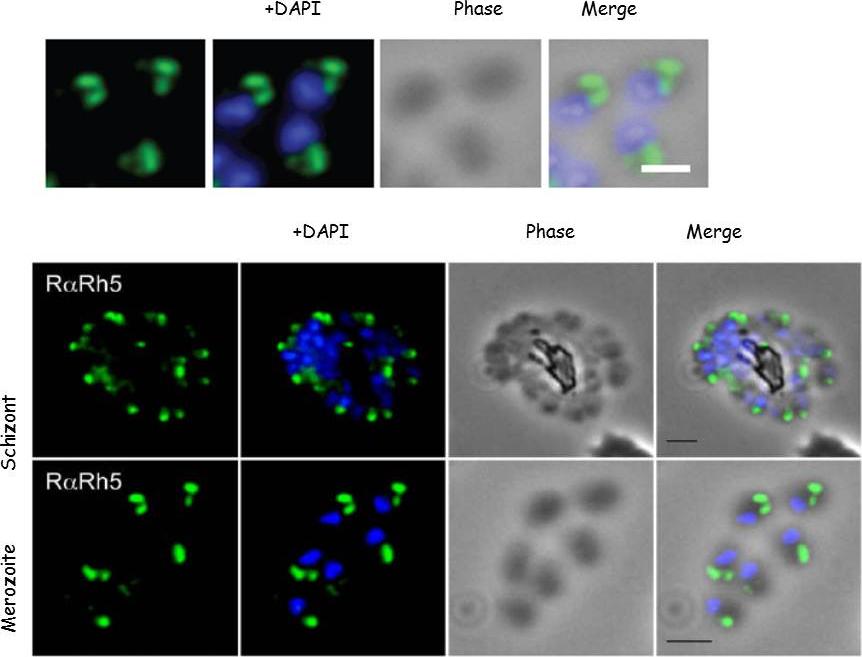
Schizont and merozoite culture was incubated overnight with 0.1µM cytochalasin D to inhibit invasion. Thin smeared on a glass slide, fixed in methanol, stained with Rabbit anti-PfRh5 polyclonal serum 1:200 and secondary Alexa-Flor488 anti-Rabbit. Dual colour fluorescence images were captured using a Carl Zeiss Axioskop 2 microscope at 100x using Oil imersion. Nucleus is marked with DAPI. Scale bar indicate 1µM. PfRh5 localizes to the rhoptries (as shown by immuno-EM).Baum J, Chen L, Healer J, Lopaticki S, Boyle M, Triglia T, Ehlgen F, Ralph SA, Beeson JG, Cowman AF. Reticulocyte-binding protein homologue 5 - An essential adhesin involved in invasion of human erythrocytes by Plasmodium falciparum. Int J Parasitol. 2008 39(3):371-80 . Copyright Elsevier 2009.
See original on MMP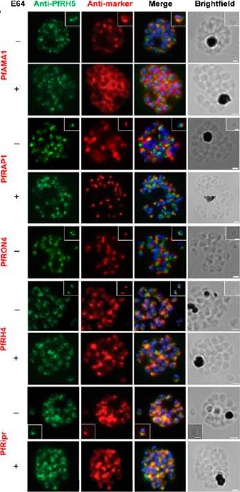
Localization of PfRH5 was assessed by indirect IFA using anti-PfRH5 polyclonal rabbit serum (green). Fixed and permeabilized schizonts with (+) or without (2) E64 treatment or free merozoites (inset) of 3D7 clone P. falciparum were costained with mouse Abs (red) to mark various organelles: PfAMA1 polyclonal (microneme), PfRAP1 mAb (rhoptry body), or PfRON4 mAb (rhoptry neck); or polyclonal mouse serum against further Ags: PfRH4 and PfRipr. Figures show the merge of the dual staining Abs and nuclei stained with DAPI (blue), as well as the brightfield view. Scale bars, 1 mm. anti-PfRH5 rabbit Abs (8) do not colocalize with conventional markers of the rhoptry bulb, rhoptry neck, or micronemes (PfRAP1, PfRON4, and PfAMA1, respectively) in permeabilized schizonts or free merozoites of 3D7 clone P. falciparum parasites. Minimal colocalization in schizonts with PfRH2a/b (data not shown) and also with PfRH4, described as a marker of the rhoptry tip, but significant colocalization with PfRipr in merozoites and late-stage schizonts, especially following treatment with the cysteine protease inhibitor E64, which prevents merozoite release by inhibiting schizont rupture. Impossible to detect PfRH5 on the merozoite surface in any assay, with staining only successful following permeabilization .Douglas AD, Williams AR, Knuepfer E, Illingworth JJ, Furze JM, Crosnier C, Choudhary P, Bustamante LY, Zakutansky SE, Awuah DK, Alanine DG, Theron M, Worth A, Shimkets R, Rayner JC, Holder AA, Wright GJ, Draper SJ. Neutralization of Plasmodium falciparum Merozoites by Antibodies against PfRH5. J Immunol. 2014 192(1):245-58.
See original on MMP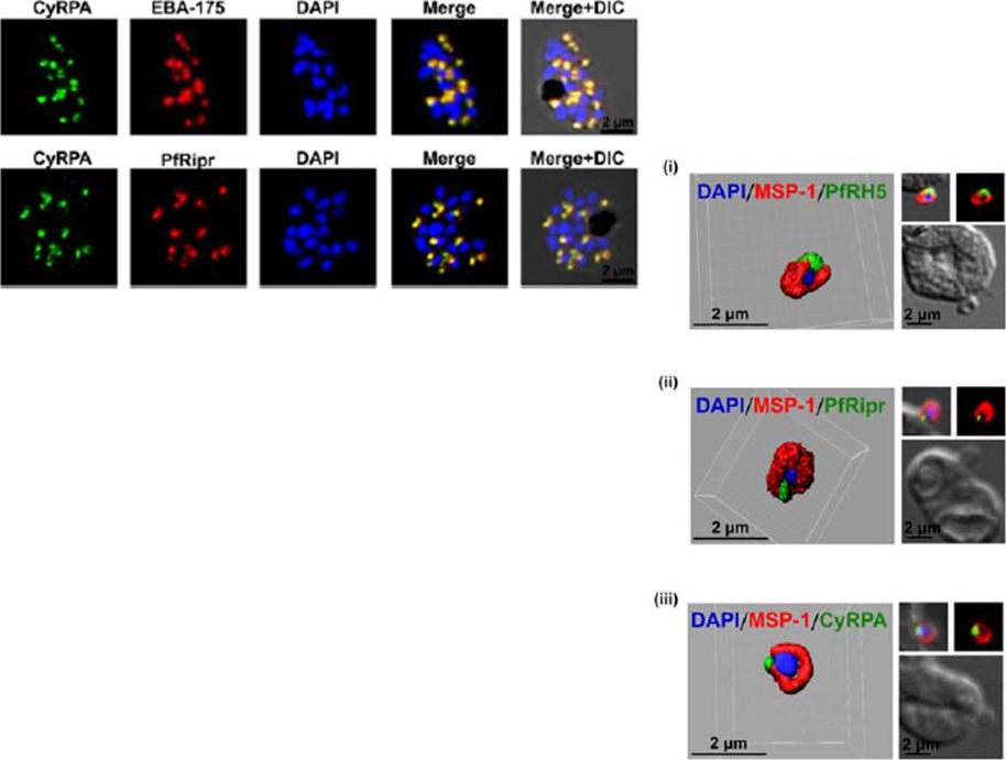
Left: Immunofluorescence microscopy detected CyRPA to colocalize with micronemal proteins (EBA-175, PfRipr). (Scale bar, 2 μm.)Right: PfRH5/PfRipr/CyRPA complex is GPI-anchored to the merozoite surface. (A) 3D reconstruction of IFA z-stacks during merozoite invasion, coimmunostained with MSP-1 detected PfRH5, PfRipr, and CyRPA at the apical end of the merozoite surface. In the 3D images, the γ settings were altered for visual representation only,Reddy KS, Amlabu E, Pandey AK, Mitra P, Chauhan VS, Gaur D. Multiprotein complex between the GPI-anchored CyRPA with PfRH5 and PfRipr is crucial for Plasmodium falciparum erythrocyte invasion. Proc Natl Acad Sci U S A. 2015 Jan 12.
See original on MMP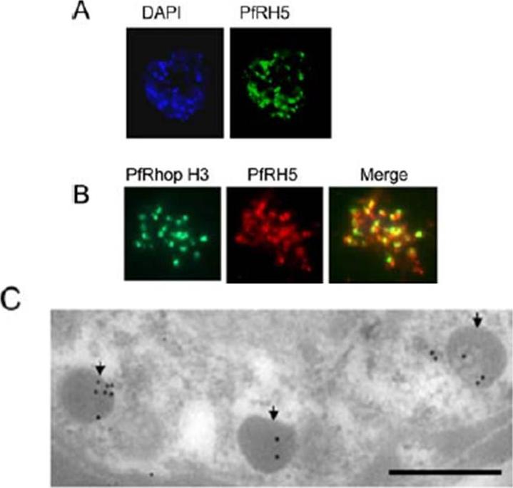
Apical localization of PfRH5 in the merozoite. A Mature Dd2 schizonts were labeled with rabbit anti-PfRH5 IgG and counterstained with FITC-labeled anti-rabbit IgG (green) using immunofluorescence microscopy. B Colocalization studies: Mature Dd2 schizonts were double-labeled with anti-PfRH5 IgG (red) and anti-RhopH3 (green) monoclonal antibody. The merged staining (yellow) by both antibodies indicates their similar location within the parasite. C Immunoelectron microscopy confirming the localization of PfRH5 in the rhoptries within the developing merozoites in the Dd2 schizont. Parasites were fixed with 1% paraformaldehyde and 0.1% glutaraldehyde. Staining was detected with rabbit anti-RH5 and antirabbit gold (10 nm). Scale bar represents 500 nm.Rodriguez M, Lustigman S, Montero E, Oksov Y, Lobo CA. PfRH5: a novel reticulocyte-binding family homolog of plasmodium falciparum that binds to the erythrocyte, and an investigation of its receptor. PLoS One. 2008 3(10):e3300.
See original on MMP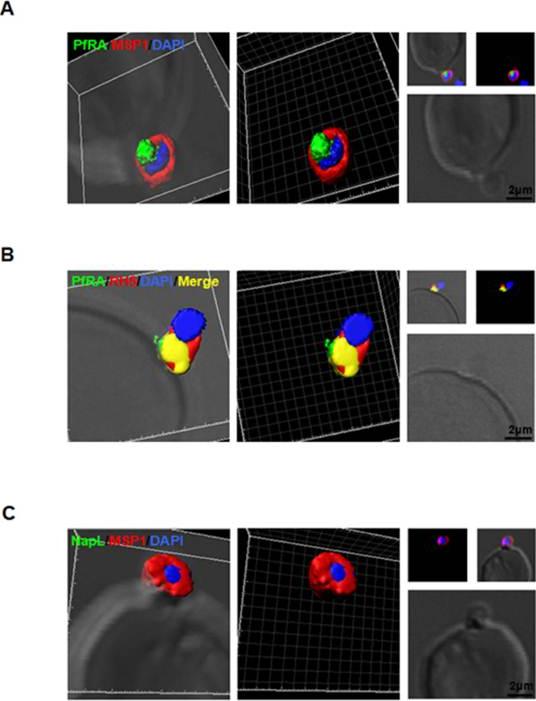
PfRA is present at the apical surface of an invading merozoite. Confocal immunofluorescencemicroscopy analysis of PfRA localization in cytochalasin-D treated invading merozoites under non-permeable fixing conditions using glutaraldehyde /paraformaldehyde. (A) 3D reconstruction of z-stacks during merozoite invasion co-immunostained with MSP-1and PfRA. PfRA detected at the apical end of the merozoite surface. In the 3D images, the γ settings were altered for visual representation only. (B) PfRA (green) co-localizes with rhoptry bulb marker PfRH5 (red) that is known to be translocated to the merozoite surface to engage with its erythrocyte receptor, Basigin. (C) As a control, staining of a nuclear parasite protein, NapL, was not detected under the same non-permeable fixing conditions confirming that the fluorescent signals were obtained only from surface localized proteins. Nuclei were stained with DNA intercalating dye DAPI (Scale bar, 2 μ m.)Anand G, Reddy KS, Pandey AK, Mian SY, Singh H, Mittal SA, Amlabu E, Bassat Q, Mayor A, Chauhan VS, Gaur D. A novel Plasmodium falciparum rhoptry associated adhesin mediates erythrocyte invasion through the sialic-acid dependent pathway. Sci Rep. 2016 6:29185.
See original on MMP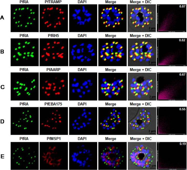
PfRA localizes to the rhoptries of P. falciparum schizonts and merozoites. Co-localization of PfRA with (A) PfTRAMP, (B) PfRH5, (C) PfAARP, (D) PfEBA-175 and (E) MSP-1 in schizont stage parasites by confocal immunoflourescence microscopy. PfRA (green) co-localizes with rhoptry bulb protein PfTRAMP(red) and PfRH5 (red) but does not co-localize with the micronemal protein PfEBA-175 (red) and merozoite surface protein MSP-1 in schizonts. PfRA (green) showed partial co-localization with rhoptry neck protein PfAARP (red). The co-localization of PfRA was quantitatively analyzed and the Pearson correlation co-efficientwith PfTRAMP (0.87) and PfRH5 (0.82) suggested that the two proteins were co-localized with PfRA. The coefficients of PfRA with PfAARP, PfEBA-175 and MSP1 were lower than 0.7 indicating that the three proteins were not co-localized with PfRA. Nuclei were stained with DNA intercalating dye DAPI. (Scale bar, 2 μm.)Anand G, Reddy KS, Pandey AK, Mian SY, Singh H, Mittal SA, Amlabu E, Bassat Q, Mayor A, Chauhan VS, Gaur D. A novel Plasmodium falciparum rhoptry associated adhesin mediates erythrocyte invasion through the sialic-acid dependent pathway. Sci Rep. 2016 6:29185.
See original on MMP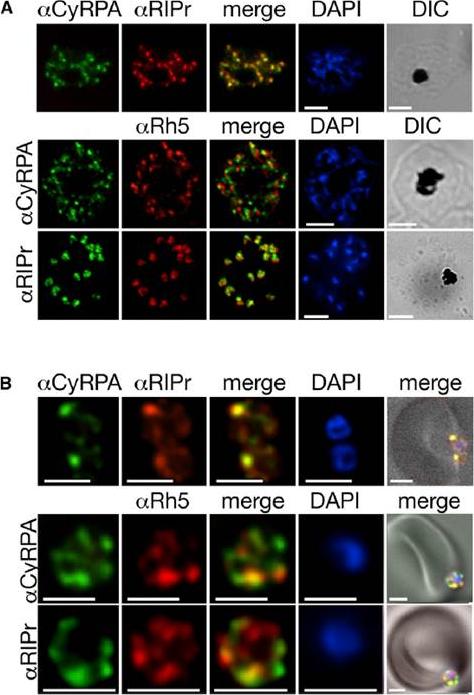
Subcellular Localization of PfRipr, CyRPA, and PfRh5 in Schizonts and Invading Merozoites(A) Top panel: Overlay of CyRPA (green) and PfRipr (red) with DAPI and DIC fields. Middle panel, overlay of CyRPA (green) and PfRh5 (red) with DAPI andDIC fields. Lower panel, overlay of PfRipr (green) and PfRh5 (red) with DAPI and DIC images.(B) Merozoites were mixed with erythrocytes and CyRPA, PfRipr, and PfRh5 visualized. Top panel, colocalization of CyRPA (green) and PfRipr (red) with DAPI and DIC images. Two merozoites invading a single erythrocyte. Middle panel, overlay of CyRPA (green) and PfRh5 (red) with DAPI and DIC fields with a single merozoite. Lower panel, overlay of PfRipr (green) and PfRh5 (red) withDAPI and DIC images. White bar indicates 5 mm.Volz JC, Yap A, Sisquella X, Thompson JK, Lim NT, Whitehead LW, Chen L, Lampe M, Tham WH, Wilson D, Nebl T, Marapana D, Triglia T, Wong W, Rogers KL, Cowman AF. Essential Role of the PfRh5/PfRipr/CyRPA Complex during Plasmodium falciparum Invasion of Erythrocytes. Cell Host Microbe. 2016 Jun 30 [Epub ahead of print] PMID: 27374406
See original on MMP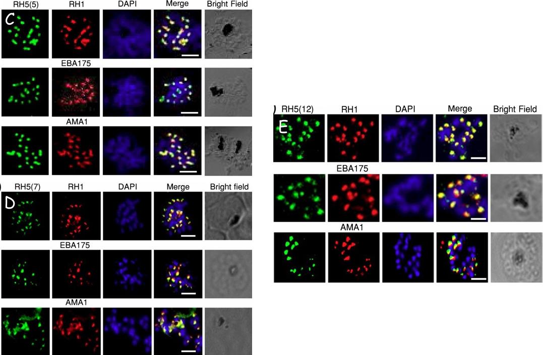
Validation of RH5 mAbs by Immunofluroscence Microscopy. Smears of 3D7 late stage schizont were co- stained with RH5(5) (c) (7) (d) and (12) (e) (first column, green) and antibodies against other invasion markers RH1, EBA175 and AMA1 respectively (second column, red). Parasite nuclei were stained with DAPI (blue). In the merged images, areas of overlap between the red and the green signals are shown in yellow. Bright field indicates the outline of the schizont. Scale bar =2μm.Aniweh Y, Gao X, Hao P, Meng W, Lai SK, Gunalan K, Chu TT, Sinha A, Lescar J, Chandramohanadas R, Li HY, Sze SK, Preiser PR. RH5-Basigin interaction induceschanges in the cytoskeleton of the host RBC. Cell Microbiol. 2017 Apr 13 [Epub ahead of print] PMID: 28409866
See original on MMPMore information
| PlasmoDB | PF3D7_0424100 |
| GeneDB | PF3D7_0424100 |
| Malaria Metabolic Pathways | Localisation images Pathways mapped to |
| Previous ID(s) | MAL4P1.224, PFD1145c |
| Orthologs | |
| Google Scholar | Search for all mentions of this gene |