PF3D7_0423700 early transcribed membrane protein 4 (ETRAMP4)
Disruptability [+]
| Species | Disruptability | Reference | Submitter |
|---|---|---|---|
| P. falciparum 3D7 |
Possible |
USF piggyBac screen (Insert. mut.) | USF PiggyBac Screen |
Mutant phenotypes [+]
None reported yet. Please press the '+' button above to add one.Imaging data (from Malaria Metabolic Pathways)
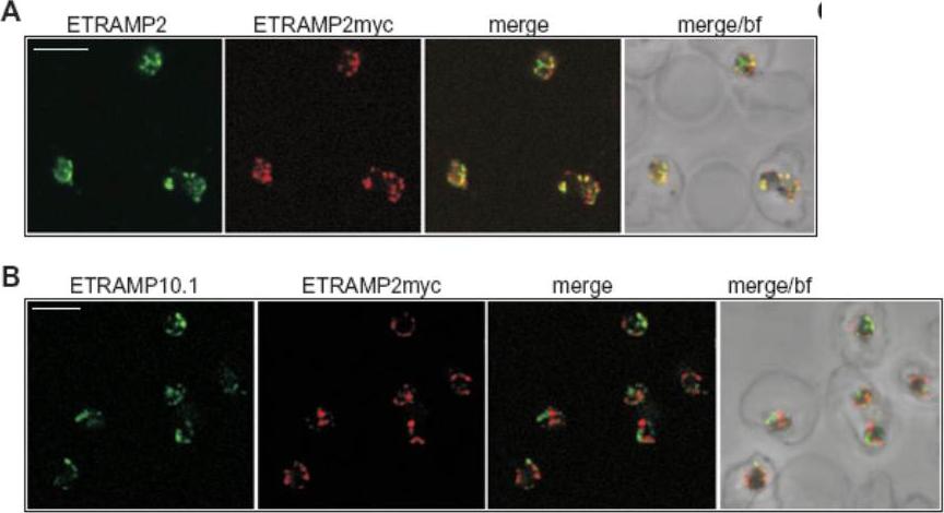
Confocal microscopy demonstrates patching of different ETRAMPs into distinct domains in formaldehyde-fixed IRBCs. IRBCs (D10E2myc parasites) coated on 10-well slides using concanavalinA (fixed with 1% formaldehyde and permeabilized with 0.03% saponin) were incubated with sera specific for ETRAMP2, ETRAMP10.1, ETRAMP2myc (detected via the tag) or anti-ETRAMP2 serum labelled with a fluorescent Fab fragment and viewed by confocal microscopy. A. ETRAMP2 (cy2, green) co-patches with ETRAMP2myc (cy3, red) into distinct areas in the PVM of several ring-stage IRBCs. B. ETRAMP10.1 (cy2, green) patches into distinct areas when compared with ETRAMP2myc (cy3, red) in several ring-stage IRBCs. bf, brightfield; bar size, 4 μm.Spielmann T, Gardiner DL, Beck HP, Trenholme KR, Kemp DJ. Organization of ETRAMPs and EXP-1 at the parasite-host cell interface of malaria parasites. Mol Microbiol. 2006 59:779-94. PMID:
See original on MMP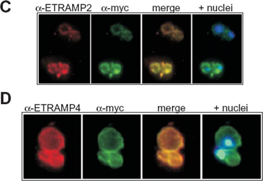
Transgenic expression of myc-tagged ETRAMP2 and ETRAMP4. Immunofluorescence analysis in acetone-fixed IRBCs: the tagged transgenes detected via the myc tag colocalize with the respective chromosomally derived ETRAMPs in a typical peripheral staining in the transfected parasites D10E2myc (C) and D10E4myc (D). C shows two IRBCs, each with a double infection of ring-stage parasites and D shows a double infection of trophozoites. Hoechst was used to stain the nuclei. Immunofluorescence analysis with an anti-myc serum showed a circular staining pattern in acetone-fixed D10E2myc and D10E4myc parasites (C and D), which is typical of the ETRAMP staining pattern seen with anti-ETRAMP serum in untransfected parasites. ETRAMPs are seen in focused areas of the PVM.Spielmann T, Gardiner DL, Beck HP, Trenholme KR, Kemp DJ. Organization of ETRAMPs and EXP-1 at the parasite-host cell interface of malaria parasites. Mol Microbiol. 2006 59:779-94.
See original on MMP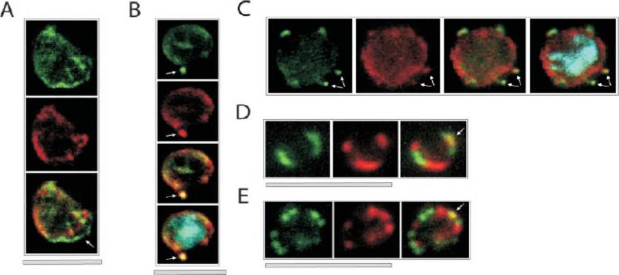
Colocalization of ETRAMPs with EXP-1 and SERP. The first image in each set shows ETRAMP labeling (green, FITC), the second Texas-Red-labeled SERP (A) or EXP-1 (B–F), and the third image represents the merged picture. The fourth panel in B and C depicts the merged picture, including nuclear staining (DAPI). (A) Confocal microscopy analysis of SERP and ETRAMP4 colocalization in schizont stage parasites (dried and 1% formaldehyde fixed). The ETRAMP4 signal seems partially detached from the PV content (arrow). (B and C) ETRAMP4 and EXP-1 localize to dots, probably representing vesicular structures that are detached from the PVM (E) or seem connected to the PVM (F). Dots are indicated by arrows. (D and E) Fluorescent microscopic analysis of ETRAMP and EXP-1 costaining of a ring-stage parasite displaying an even circular pattern (D, 0.1% formaldehyde-fixed IRBCs, ETRAMP10.1) or a beads on a string pattern (E, unfixed IRBCs, ETRAMP2). Yellow color shows regions of overlap (arrows). This indicated that ETRAMPs and EXP-1 localize to different regions of the PVM.Spielmann T, Fergusen DJ, Beck HP. etramps, a new Plasmodium falciparum gene family coding for developmentally regulated and highly charged membrane proteins located at the parasite-host cell interface. Mol Biol Cell. 2003 14:1529-44.
See original on MMP
ETRAMPs are located in the PVM with their C termini facing the RBC cytosol. (A–C) Distribution of ETRAMP4 around the parasite periphery in fixed cells. Top two panels of A depict ETRAMP4 staining (FITC) and nuclear staining (DAPI) of a schizont stage parasite; spots of more intense staining are indicated by small arrows. Bottom two panels of A show ETRAMP4 (FITC) and nuclear staining (DAPI) of a late trophozoite stage parasite. The single patch of strong staining is indicated by a large arrow. (B) Extensions from the parasite body are labeled by anti-ETRAMP4 serum, indicating localization in blebbing vesicles or the TVM. The top panel shows ETRAMP4 (FITC) detected in a young trophozoite; below the nucleus is shown (DAPI). The extension is indicated by an arrow. (C) Costaining with a rabbit anti-aldolase serum verifies that the periphery of the parasite is decorated by ETRAMP4 antiserum; the four panels represent the same section depicting a young trophozoite. From top to bottom, ETRAMP4 (FITC), aldolase (Cy3), nucleus (DAPI), and aldolase and ETRAMP4 merged. Bars, 5 mm.Spielmann T, Fergusen DJ, Beck HP. etramps, a new Plasmodium falciparum gene family coding for developmentally regulated and highly charged membrane proteins located at the parasite-host cell interface. Mol Biol Cell. 2003 14:1529-44.
See original on MMP
Upper row: Location of REX3 in IRBCs fixed with 4% formaldehyde/0.005% glutaraldehyde. IFAs with 3D7 parasites expressing a myc-tagged ETRAMP4 showed that REX3 (red; Texas Red) occurs as a uniform stain outside of the PVM (ETRAMP4myc in the PVM detected by a rabbit anti-myc antibody; green, Cy2). Second row: REX3 (red; Texas Red) did not colocalize with the Maurer’s cleft protein REX1 (green; Cy2) in 3D7 parasites. Bars, 5 mm.Lower panel: Confocal fluorescence microscopy images of transfected 3D7 P. falciparum-infected RBCs. REX3 is expressed in its early ring stages (top row) and is directed to the bulk compartment of the host cell cytoplasm. Trophozoites and schizonts (middle and bottom rows) display a uniform fluorescence pattern with some puncta. Tilley L, McFadden G, Cowman A, Klonis N. Illuminating Plasmodium falciparum-infected red blood cells. Trends Parasitol. 2007 23:268-77.
See original on MMPMore information
| PlasmoDB | PF3D7_0423700 |
| GeneDB | PF3D7_0423700 |
| Malaria Metabolic Pathways | Localisation images Pathways mapped to |
| Previous ID(s) | MAL4P1.219, PFD1120c |
| Orthologs | |
| Google Scholar | Search for all mentions of this gene |