PF3D7_0423400 apical asparagine-rich protein AARP (AARP)
Disruptability [+]
No information reported yet. Please press the '+' button above to add some.Mutant phenotypes [+]
None reported yet. Please press the '+' button above to add one.Imaging data (from Malaria Metabolic Pathways)
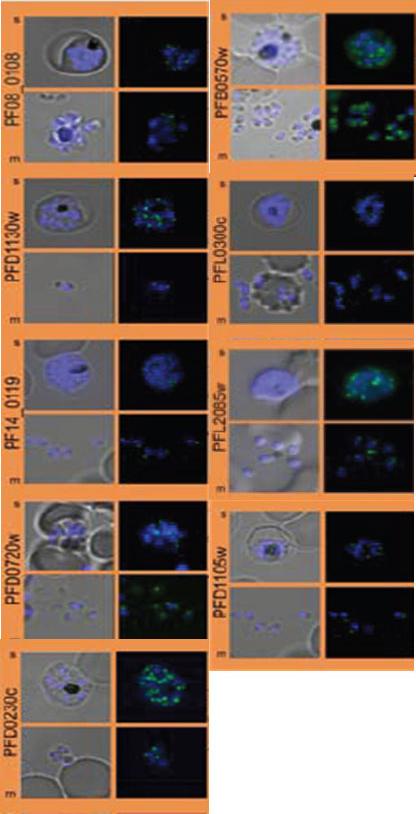
Subcellular distribution of proteins predicted to be involved in invasion. All proteins were localized in schizonts (s) and free merozoites (m) using GFP-fusion proteins and grouped according to their predominant GFP localization. Apical. Nuclei were stained with DAPI (blue).Hu G, Cabrera A, Kono M, Mok S, Chaal BK, Haase S, Engelberg K, Cheemadan S, Spielmann T, Preiser PR, Gilberger TW, Bozdech Z. Transcriptional profiling of growth perturbations of the human malaria parasite Plasmodium falciparum. Nat Biotechnol. 2010 28(1):91-8.
See original on MMP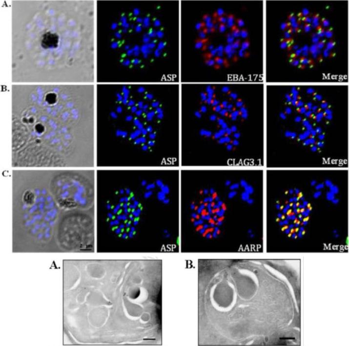
Co-localization of PfASP in late stage P. falciparum schizonts with microneme protein, EBA-175 (A); rhoptry bulb protein, Clag3.1 (B); and rhoptry neck protein, PfAARP (C). P. falciparum schizonts were stained with anti-PfASP-RIV mouse sera (green) and either anti-EBA-175 (red), anti-CLAG3.1 (red) or anti PfAARP (red) rabbit sera. Parasite nuclei were stained with DAPI (blue). PfASP did not co-localize with microneme protein EBA-175 or rhoptry bulb protein CLAG3.1 but co-localized with rhoptry neck protein PfAARP in segmented P. falciparum merozoites within late-stage schizonts. Bottom: Ultra thin sections of late-stage P. falciparum schizonts containing segmented merozoites were labelled with anti-PfASP-RIV mouse sera and gold-conjugated anti-mouse IgG. Labelling was observed in the neck of rhoptries in segmented merozoites of late-stage P. falciparum schizonts. Scale bar = 250 nm.Srivastava A, Singh S, Dhawan S, Alam MM, Mohmmed A, Chitnis CE. Localization of apical Sushi Protein in Plasmodium falciparum merozoites. Mol Biochem Parasitol. 2010 74(1):66-9 Copyright Elsevier2011
See original on MMP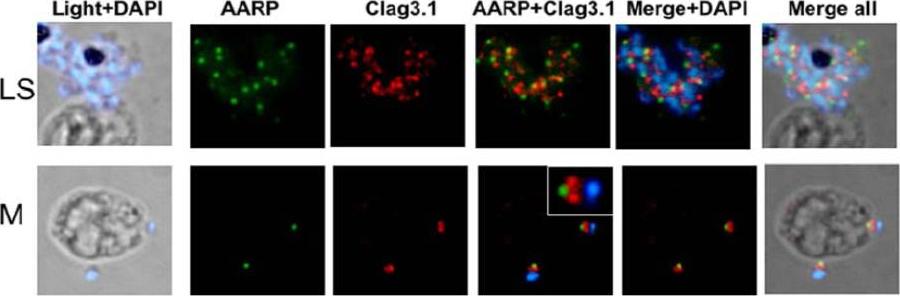
Spatial localization of PfAARP by co-immunostaining studies with rhoptry resident protein Clag3.1. P. falciparum parasites were coimmuno-stained with anti- PfAARP (green) and anti-Clag3.1 (red) antibodies. The parasite nuclei were stained with DAPI (blue). Clag3.1 staining was present in the rhoptry bulb in the free and invading merozoites (lower panel). PfAARP was localized just above these two rhoptry bulbs towards the apex of the merozoites. Enlarged image of merozoite invading in the host erythrocyte is shown in the insetWickramarachchi T, Devi YS, Mohmmed A, Chauhan VS. Identification and characterization of a novel Plasmodium falciparum merozoite apical protein involved in erythrocyte binding and invasion. PLoS ONE. 2008 Mar 5;3(3):e1732.
See original on MMP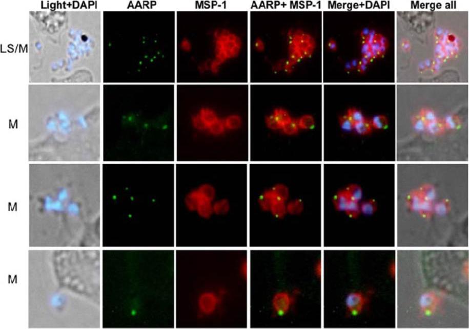
Immunofluorescence assay to localize PfAARP by coimmuno-staining of P. falciparum parasites with anti-PfAARP (green) and anti-MSP-1 (red) antibodies. The parasite nuclei were stained with DAPI (blue) and slides were visualized by fluorescence microscope. The apical ends of the merozoites have dense structure. MSP-1 staining was found around the merozoites and the PfAARP was localized at the apex of the merozoites.Wickramarachchi T, Devi YS, Mohmmed A, Chauhan VS. Identification and characterization of a novel Plasmodium falciparum merozoite apical protein involved in erythrocyte binding and invasion. PLoS ONE. 2008 Mar 5;3(3):e1732.
See original on MMP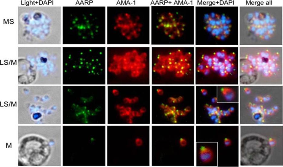
Spatial localization of merozoite’s microneme resident proteins AMA-1 and PfAARP by co-immunostaining with anti-PfAARP (green) and anti-AMA-1 (red) Abs: both showed punctate staining in the schizonts. In the late schizonts and merozoites, AMA-1 was present over the entire surface of the merozoites but is most densely distributed at their apical tip. Enlarged image of selected individual merozoite is shown in the inset. Wickramarachchi T, Devi YS, Mohmmed A, Chauhan VS. Identification and characterization of a novel Plasmodium falciparum merozoite apical protein involved in erythrocyte binding and invasion. PLoS ONE. 2008 Mar 5;3(3):e1732.
See original on MMP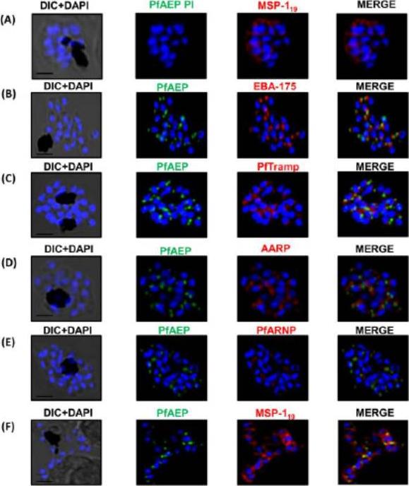
PfAEP localization in schizont stages analyzed by confocal microscopy. Subcellular localization of PfAEP was studied by co-staining with antibodies against micronemal protein EBA-175 (B), rhoptry bulb protein PTRAMP (C), and rhoptry neck,apical asparagine rich protein (AARP) (D), apical rhoptry neck protein (ARNP) (E), and merozoite surface protein (MSP) (F).As a control, pre-immune serum of PfAEP was checked and no staining was observed in schizonts (A). Mature schizonts were costained with anti-PfAEP (green) and anti-EBA-175, TRAMP, AARP, ARNP, MSP-119 antibodies (red). The nuclei of schizonts were stained with DAPI (blue) and visualized by Nikon N-SIM confocal microscope. All apical marker proteins and PfAEP showed punctate staining in schizonts distinct from DAPI. Co-staining of PfAEP with surface marker MSP-119 showed PfARNP localized at the apical tip with surface of merozoites stained by MSP-119. PfAEP staining did not merge with either markers of microneme or rhoptry indicating that PfAEP is neither a resident of micronemes or rhoptry ofmerozoites. Scale bar 2 mm.Hans N, Relan U, Dubey N, Gaur D, Chauhan VS. Identification and localization of a Novel Invasin of Plasmodium falciparum. Mol Biochem Parasitol. 2015 Sep 29. [Epub ahead of print]
See original on MMP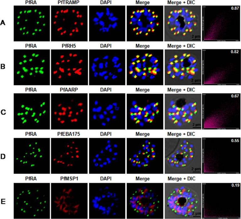
PfRA localizes to the rhoptries of P. falciparum schizonts and merozoites. Co-localization of PfRA with (A) PfTRAMP, (B) PfRH5, (C) PfAARP, (D) PfEBA-175 and (E) MSP-1 in schizont stage parasites by confocal immunoflourescence microscopy. PfRA (green) co-localizes with rhoptry bulb protein PfTRAMP(red) and PfRH5 (red) but does not co-localize with the micronemal protein PfEBA-175 (red) and merozoite surface protein MSP-1 in schizonts. PfRA (green) showed partial co-localization with rhoptry neck protein PfAARP (red). The co-localization of PfRA was quantitatively analyzed and the Pearson correlation co-efficientwith PfTRAMP (0.87) and PfRH5 (0.82) suggested that the two proteins were co-localized with PfRA. The coefficients of PfRA with PfAARP, PfEBA-175 and MSP1 were lower than 0.7 indicating that the three proteins were not co-localized with PfRA. Nuclei were stained with DNA intercalating dye DAPI. (Scale bar, 2 μm.)Anand G, Reddy KS, Pandey AK, Mian SY, Singh H, Mittal SA, Amlabu E, Bassat Q, Mayor A, Chauhan VS, Gaur D. A novel Plasmodium falciparum rhoptry associated adhesin mediates erythrocyte invasion through the sialic-acid dependent pathway. Sci Rep. 2016 6:29185.
See original on MMPMore information
| PlasmoDB | PF3D7_0423400 |
| GeneDB | PF3D7_0423400 |
| Malaria Metabolic Pathways | Localisation images Pathways mapped to |
| Previous ID(s) | MAL4P1.216, PFD1105w |
| Orthologs | PBANKA_0523800 , PCHAS_0524000 , PKNH_0515300 , PVP01_0531900 , PVX_090210 , PY17X_0525200 |
| Google Scholar | Search for all mentions of this gene |