PF3D7_0417200 bifunctional dihydrofolate reductase-thymidylate synthase (DHFR-TS)
Disruptability [+]
| Species | Disruptability | Reference | Submitter |
|---|---|---|---|
| P. falciparum 3D7 |
Refractory |
USF piggyBac screen (Insert. mut.) | USF PiggyBac Screen |
Mutant phenotypes [+]
| Species | Stage | Phenotype | Reference | Submitter |
|---|---|---|---|---|
| P. falciparum 3D7 | Asexual |
Attenuated |
24023691 (Knock down) | Theo Sanderson, Francis Crick Institute |
Imaging data (from Malaria Metabolic Pathways)
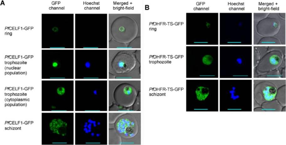
Protein localization of PfCELF1 and PfDHFR-TS. PfCELF1 was expressed as a fusion protein with C-terminal GFP in a transgenic parasite line established in 3D7 strain. The integrant transgenic line clone #1 expressing GFP-tagged PfDHFR-TS established in 3D7 strain for comparison was described previously [36]. (A) Representative confocal microscopic images of transgenic PfCELF1-GFP parasites. Images are shown for each asexual developmental stage (ring, trophozoite and schizont); left panel is the GFP signal showing PfCELF1-GFP, middle panel is Hoechst signal showing nucleus and right panel is the merge of both fluorescent signals overlaid with the bright-field image. Scale bars, 5 mm. (B) Representative confocal microscopic images of transgenic PfDHFR-TS-GFP parasites. Images are shown for each asexual developmental stage and arranged as in part A. Scale bars, 5 mm. Wongsombat C, Aroonsri A, Kamchonwongpaisan S, Morgan HP, Walkinshaw MD, Yuthavong Y, Shaw PJ. Molecular characterization of Plasmodium falciparumBruno/CELF RNA binding proteins. Mol Biochem Parasitol. 2014 Nov 3;198(1):1-10. [Epub ahead of print]
See original on MMP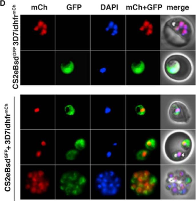
Communication between Parasites Results in the DNA-Dependent Transfer of Drug Resistance and Fluorescence.. Fluorescence microscopy of live 3D7idhfrmCh (mCherry) and CS2eBsdGFP (GFP) cocultured. Top, 3D7idhfrmCh expression of mCherry (red) in nucleus. Second panel, CS2eBsdGFP expression of GFP in cytoplasm. Bottom, examples of CS2eBsdGFP+3D7idhfrmCh cocultures in Bs+WR after 5 days. DAPI-stained images (blue) show the nucleus and final image merged.Regev-Rudzki N, Wilson DW, Carvalho TG, Sisquella X, Coleman BM, Rug M, Bursac D, Angrisano F, Gee M, Hill AF, Baum J, Cowman AF. Cell-Cell Communication between Malaria-Infected Red Blood Cells via Exosome-like Vesicles. Cell. 2013 May 23;153(5):1120-33.
See original on MMPMore information
| PlasmoDB | PF3D7_0417200 |
| GeneDB | PF3D7_0417200 |
| Malaria Metabolic Pathways | Localisation images Pathways mapped to |
| Previous ID(s) | MAL4P1.161, PFD0830w |
| Orthologs | PBANKA_0719300 , PCHAS_0728300 , PKNH_0509600 , PVP01_0526600 , PVX_089950 , PY17X_0719300 |
| Google Scholar | Search for all mentions of this gene |