PF3D7_0414900 armadillo-domain containing rhoptry protein (ARO)
Disruptability [+]
No information reported yet. Please press the '+' button above to add some.Mutant phenotypes [+]
None reported yet. Please press the '+' button above to add one.Imaging data (from Malaria Metabolic Pathways)
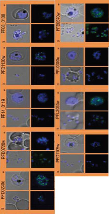
Subcellular distribution of proteins predicted to be involved in invasion. All proteins were localized in schizonts (s) and free merozoites (m) using GFP-fusion proteins and grouped according to their predominant GFP localization. Apical. Nuclei were stained with DAPI (blue).Hu G, Cabrera A, Kono M, Mok S, Chaal BK, Haase S, Engelberg K, Cheemadan S, Spielmann T, Preiser PR, Gilberger TW, Bozdech Z. Transcriptional profiling of growth perturbations of the human malaria parasite Plasmodium falciparum. Nat Biotechnol. 2010 28(1):91-8.
See original on MMP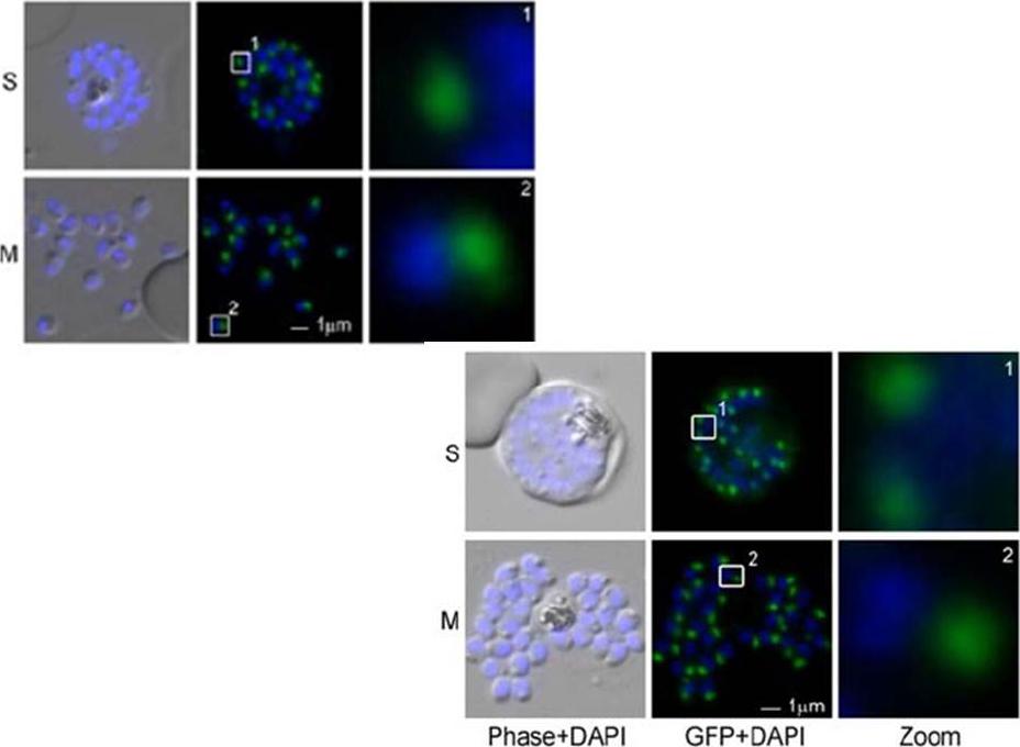
Upper left: PfARO rhoptry membrane association. Nuclei stained with DAPI (blue). Enlargement of selected areas are marked with white square and referred as Zoom. Scale bar, 1 μm.Lower right: The N-terminus of ARO is sufficient for rhoptry association and is palmitoylated. (A) Schematic representation of the minimal PfARO construct (20AROwt) encompassing the N-terminal 20 amino acids fused to GFP and its apical localization unfixed parasites (S, schizonts and M, merozoites). Nuclei stained with DAPI (blue). Enlargement of selected areas are marked with white square and referred as Zoom. Scale bar, 1 μm. Cabrera A, Herrmann S, Warszta D, Santos JM, John Peter AT, Kono M, Debrouver S, Jacobs T, Spielmann T, Ungermann C, Soldati-Favre D, Gilberger TW. Dissection of minimal sequence requirements for rhoptry membrane targeting in the malaria parasite. Traffic. 2012 13(10):1335-50.
See original on MMP
ARO is late transcribed in blood stages of Plasmodium falciparum and co-localizes with the rhoptries. Co-localization of PfARO with (upper panel) the rhoptry marker RALP-1 (red) and (lower panel) the microneme marker EBA-181 (red). Nuclei stained with DAPI (blue). Enlargement of selected areas are marked with white square and referred as Zoom. Scale bar, 1 μm. Colocalization with the rhoptry bulb marker RALP-1 and the microneme marker EBA-181 identify PfARO as a rhoptry protein.Cabrera A, Herrmann S, Warszta D, Santos JM, John Peter AT, Kono M, Debrouver S, Jacobs T, Spielmann T, Ungermann C, Soldati-Favre D, Gilberger TW. Dissection of minimal sequence requirements for rhoptry membrane targeting in the malariaparasite. Traffic. 2012 13(10):1335-50. PMID:
See original on MMP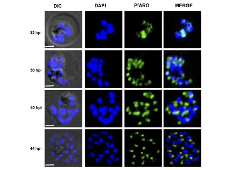
Native PfARO exhibits nucleo-apical shuttling. Confocal immunofluorescence imaging using PfARO specific antibodies detected predominant expression of native PfARO in the nucleus during the early schizont stage parasites, which decreased with the intra-erythrocytic growth of the parasite. At later stages, native PfARO was observed to be predominantly outside the nucleus (Scale bar, 2 μm).Mitra P, Gupta ED, Sahar T, Pandey AK, Dangi P, Reddy KS, Chauhan VS, Gaur D. Evidence for the Nucleo-Apical Shuttling of a Beta-Catenin Like Plasmodium falciparum Armadillo Repeat Containing Protein. PLoS One. 2016 11(2):e0148446.
See original on MMPMore information
| PlasmoDB | PF3D7_0414900 |
| GeneDB | PF3D7_0414900 |
| Malaria Metabolic Pathways | Localisation images Pathways mapped to |
| Previous ID(s) | MAL4P1.139, PFD0720w |
| Orthologs | PBANKA_0716900 , PCHAS_0726000 , PKNH_0506500 , PVP01_0521400 , PVX_089700 , PY17X_0717100 |
| Google Scholar | Search for all mentions of this gene |