PF3D7_0406200 sexual stage-specific protein precursor (Pfs16)
Disruptability [+]
| Species | Disruptability | Reference | Submitter |
|---|---|---|---|
| P. falciparum 3D7 |
Refractory |
USF piggyBac screen (Insert. mut.) | USF PiggyBac Screen |
Mutant phenotypes [+]
| Species | Stage | Phenotype | Reference | Submitter |
|---|---|---|---|---|
| P. falciparum 3D7 | Gametocyte |
Attenuated |
14698439 (Knock down) | Theo Sanderson, Wellcome Trust Sanger Institute |
Imaging data (from Malaria Metabolic Pathways)

Immunofluorescence assays, using specific antibodies, detected the proteins of interest (in green) in asexual blood stage (ABS) parasites, using MSP1 (in red). Nuclei were highlighted by Hoechst nuclear stain (in blue). Bar, 5 μm. Ngwa CJ, Scheuermayer M, Mair GR, Kern S, Brügl T, Wirth CC, Aminake MN, Wiesner J, Fischer R, Vilcinskas A, Pradel G. Changes in the transcriptome of the malaria parasite Plasmodium falciparum during the initial phase of transmission from the human to the mosquito. BMC Genomics. 2013 Apr 15;14:256
See original on MMP
PfGECO is external to Pfs16 and internal to MESA. Co-localization IFA was performed on gametocyte stages I-IV using anti-PfGECO serum (red) and either (A) anti-Pfs16 (green) serum or (B) anti-MESA (green) serum. Also shown is the parasite nucleus stained with DAPI (blue), the bright field (BF) and the merged images.Morahan BJ, Strobel C, Hasan U, Czesny B, Mantel PY, Marti M, Eksi S, Williamson KC. Functional analysis of the exported type IV HSP40 protein PfGECO in P. falciparum gametocytes. Eukaryot Cell. 2011 10(11):1492-503.
See original on MMP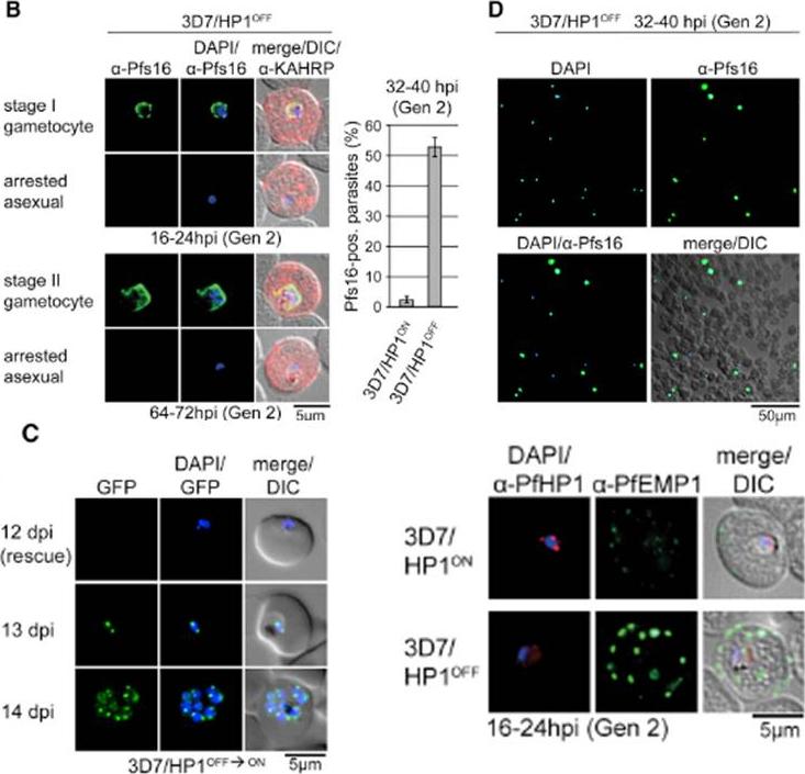
PfHP1 depletion induces gametocyte conversion. Distinction between 3D7/HP1OFF early gametocytes and arrested trophozoites by IFA (left) and proportion of Pfs16/KAHRP-positive parasites in 3D7/HP1ON and 3D7/HP1OFF (right). Values show the mean ± SD of three biological replicates (100 KAHRP-pos. iRBCs were scored per experiment). (D) a-Pfs16 IFA of a 3D7/HP1OFF parasite culture (Shield-1 removal at 4–12 hpi) at 32–40 hr postreinvasion (image taken at 40X magnification). The gametocyte hyperinduction phenotype is highlighted by the high proportion of Pfs16-positive stage I gametocytes among all DAPI-positive iRBCs. (C) Growth-arrested 3D7/HP1OFF parasites reenter mitotic proliferation after Shield-1 replenishment. dpi, days postreinvasion. PfHP1 depletion causes reversible cell-cycle arrest at the G1/S transition phase. Lower right panel: a-PfHP1/a-PfEMP1 (mAb 6H1) IFAs of 3D7/HP1ON and 3D7/HP1OFF parasites at 16–24 hpi in generation 2. PfEMP1 in 3D7/HP1OFF parasites at the single-cell level is hyperexpressed and correctly trafficked to the iRBC surfaceBrancucci NM, Bertschi NL, Zhu L, Niederwieser I, Chin WH, Wampfler R, Freymond C, Rottmann M, Felger I, Bozdech Z, Voss TS. Heterochromatin protein 1 secures survival and transmission of malaria parasites. Cell Host Microbe. 2014 16(2):165-76.
See original on MMP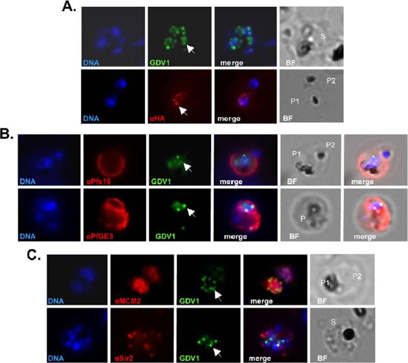
Subcellular localization of PfGDV1. Parasites transformed with GFP- or HA-tagged PfGDV1 were stained with DAPI (DNA stain) and the indicated anti-sera, and then examined by fluorescence microscopy. Images are shown of the DAPI stain (DNA), GFP-tagged PfGDV1 epifluorescence (GDV1), and antibodies specific for HA (aHA), Pfs16 (aPfs16), PfGE3 (aPfGE3), PfMCM2 (aMCM2), and PfSir2 (aSir2). The corresponding merged and bright field (BF) images are included on the right. PfGDV1 expression is indicated with an arrow; locations of parasites in the BF image are indicated with a P for parasite or S for schizont. A) A schizont (S) (Upper) expressing GDV1 and a doubly infected erythrocyte (Lower) with one parasite (P1) expressing HA-tagged PfGDV1 (aHA) and another negative (P2) for anti-HA antibodies. B) Costaining of parasites expressing GDV1 with early gameto-cytogenesis markers. A doubly infected erythrocyte (Upper) with one parasite (P1) positive for GDV1 and aPfs16 and the other (P2) negative for both. An erythrocyte (Lower) infected with a parasite (P) positive for GDV1 and aPfGE3. C) Colocalization of PfGDV1 with nuclear proteins. A doubly infected erythrocyte (Upper) with one parasite in the plane of the image (P1) and the other below (P2). Both P1 and P2 are positive for GDV1 and aMCM2. A schizont (S) (Lower) expressing GDV1 stained with aSir2.Eksi S, Morahan BJ, Haile Y, Furuya T, Jiang H, Ali O, Xu H, Kiattibutr K, Suri A, Czesny B, Adeyemo A, Myers TG, Sattabongkot J, Su XZ, Williamson KC. Plasmodium falciparum gametocyte development 1 (Pfgdv1) and gametocytogenesis early gene identification and commitment to sexual development. PLoS Pathog. 2012;8(10):e1002964.
See original on MMP
c Immunofluorescence microscopy of sexually induced PfGEXP5-GFP transfectants (14 h pi), probed with rabbit anti-GFP (green) and mouse anti-Pfs16 (red) antibodies. PfGEXP5-GFP is expressed and exported to the host cell cytoplasm in Pfs16-positive cells (c) d Immuno-fluorescence microscopy of sexually induced PfGEXP5-GFP transfectants (14 h pi, top) and an asexual trophozoite from the same line (bottom), probed with anti-GFP (green) and anti-KAHRP (red) antibodies. Scale bars 3 μm. PfGEXP5-GFP positive cells did not exhibit KAHRP labelling at the RBC surface, while KAHRP-positive cells (i.e., asexual stages) were negative for exported PfGEXP5-GFP (d).Tibúrcio M, Dixon MW, Looker O, Younis SY, Tilley L, Alano P. Specific expression and export of the Plasmodium falciparum Gametocyte EXported Protein-5 marks the gametocyte ring stage. Malar J. 2015 14:334.
See original on MMPMore information
| PlasmoDB | PF3D7_0406200 |
| GeneDB | PF3D7_0406200 |
| Malaria Metabolic Pathways | Localisation images Pathways mapped to |
| Previous ID(s) | MAL4P1.61, PFD0310w |
| Orthologs | PKNH_0304300 , PVP01_0305600 , PVX_000930 |
| Google Scholar | Search for all mentions of this gene |