PF3D7_0405900 apical sushi protein (ASP)
Disruptability [+]
| Species | Disruptability | Reference | Submitter | |
|---|---|---|---|---|
| P. falciparum 3D7 |
Possible |
USF piggyBac screen (Insert. mut.) | USF PiggyBac Screen | |
| P. berghei ANKA |
Possible |
PlasmoGEM (Barseq) | PlasmoGEM | |
Mutant phenotypes [+]
| Species | Stage | Phenotype | Reference | Submitter |
|---|---|---|---|---|
| P. berghei ANKA | Asexual |
Attenuated |
PlasmoGEM (Barseq) | PlasmoGEM |
Imaging data (from Malaria Metabolic Pathways)

PFF0645c is released from the rhoptries only after completion of merozoite invasion. (A) IEM of PFF0645c-HA merozoites pre and post-erythrocyte invasion labeled with immunogold anti-HA (white arrows). Scale bar = 0.2 mm. (B) Widefield 3D imaging of PFF0645c-HA merozoites labeled with anti-HA, anti-PfRON4 and DAPI showing early, mid and late invasion events. Scale bar = 5 mm. 3D reconstruction with 0.2 mm grid intervals. (C) Widefield 3D imaging of PFF0645c-HA early rings (,10 min post-invasion) labeled with anti-HA, anti-PfRON4 (,PVM) anti-RAP1 (PV) or anti-MSP1 (plasma membrane) and DAPI. Scale bar = 5 mm. 3D reconstruction with 0.2 mm grid intervals.Zuccala ES, Gout AM, Dekiwadia C, Marapana DS, Angrisano F, Turnbull L, Riglar DT, Rogers KL, Whitchurch CB, Ralph SA, Speed TP, Baum J. Subcompartmentalisation of Proteins in the Rhoptries Correlates with Ordered Events of Erythrocyte Invasion by the Blood Stage Malaria Parasite. PLoS One. 2012;7(9):e46160.
See original on MMP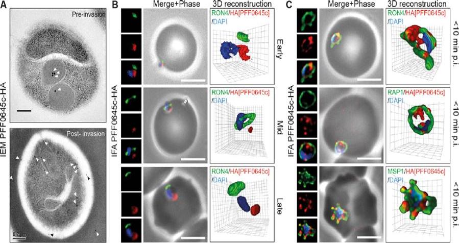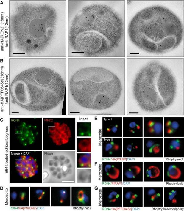
Spatial localisation of different rhoptry proteins before and during merozoite invasion. (A) IEM of free PfRON2-HA merozoites (pre-invasion) dual labeled with immunogold anti-HA (18 nm) and rhoptry bulb marker RAP1 (12 nm). Scale bar = 0.2 mm. (B) IEM of free PFF0645c-HA merozoites (pre-invasion) dual labeled with immunogold anti-HA (18 nm) and rhoptry bulb marker RAP1 (12 nm). Scale bar = 0.2 mm. (C) Widefield IFA of E64-treated schizonts (to prevent egress labeled with anti-PfRh2, anti-PfRON4 and DAPI. Scale bar = 5 mm. (D–G) Independent replicate imaging of merozoites from (D) PfRON2-HA, (E) PfASP-HA (two classes of distribution seen), (F) RAP1 and (G) PFF0645c-HA mid-way through invasion colabeled with anti-PfRON4 and DAPI. RON2-HA is located anterior to the bulb marker RAP1. PFF0645c had a more posterior or contiguous localisation to RAP1, frequently located towards the peripheral regions of the rhoptry.Zuccala ES, Gout AM, Dekiwadia C, Marapana DS, Angrisano F, Turnbull L, Riglar DT, Rogers KL, Whitchurch CB, Ralph SA, Speed TP, Baum J. Subcompartmentalisation of Proteins in the Rhoptries Correlates with Ordered Events of Erythrocyte Invasion by the Blood Stage Malaria Parasite. PLoS One. 2012;7(9):e46160.
See original on MMP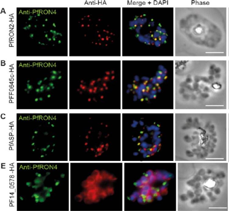
Cellular localisation of invasins in P. falciparum schizonts. (A–E) IFA of schizonts from HA tagged P. falciparum parasite lines labeled with anti-PfRON4, to mark the rhoptry neck, DAPI, to mark nuclei and anti-HA. (A) PfRON2 (B) rhoptry protein PFF0645c (C) apical sushi protein PfASP and (E) inner membrane complex protein PF14_0578. PfRON2-HA schizonts were colabeled with anti-PfRON4, a marker of the rhoptry neck. PFF0645c and PfASP each displayed a similar apical localization in schizonts, showing overlap of labeling with PfRON4. PF14_0578-HA labeling was peripheral in late stage schizonts (E), suggestive of an inner-membrane complex (IMC) or plasma membrane localization.Zuccala ES, Gout AM, Dekiwadia C, Marapana DS, Angrisano F, Turnbull L, Riglar DT, Rogers KL, Whitchurch CB, Ralph SA, Speed TP, Baum J. Subcompartmentalisation of Proteins in the Rhoptries Correlates with Ordered Events of Erythrocyte Invasion by the Blood Stage Malaria Parasite. PLoS One. 2012;7(9):e46160.
See original on MMP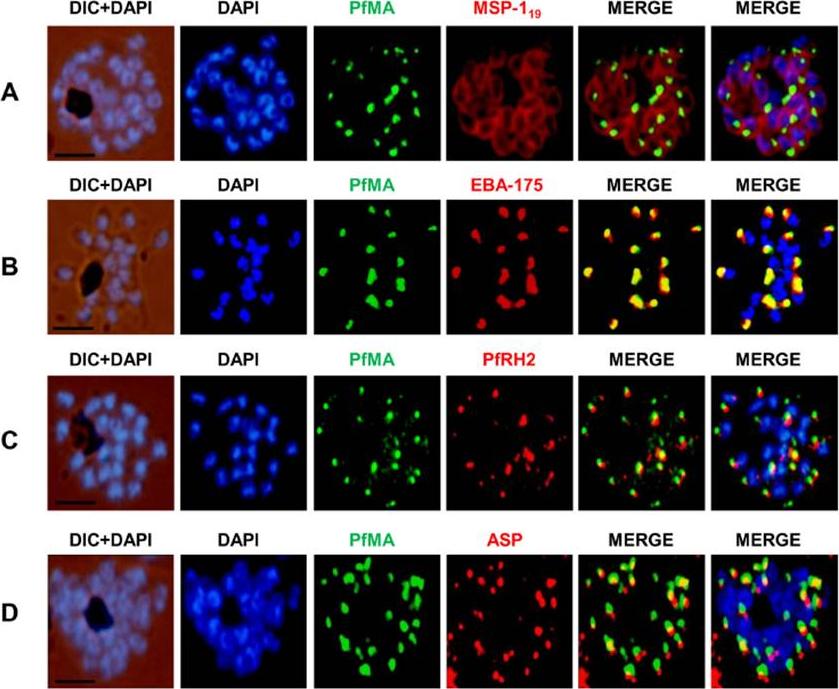
PfMA localization in schizont stages analyzed by fluorescence microscopy. Sub-cellular localization of PfMA was studied by co-immunostaining with surface protein (A), microneme (B), rhoptry (C, D) resident proteins. P. falciparum schizonts were co-immunostained with mouse anti-PfMA (green) and rabbit antibodies against one of the 4 marker proteins (EBA175/PfRH2, ASP, MSP-119) (red). The nuclei of schizont were stained with DAPI (blue) and slides were visualized by fluorescence microscope. All apical marker proteins and PfMA showed punctate staining in schizonts. PfMA was localized in the micronemes as it signal costained with micronemal marker EBA-175. The scale bar indicates 2 μm.Hans N, Singh S, Pandey AK, Reddy KS, Gaur D, Chauhan VS. Identification and Characterization of a Novel Plasmodium falciparum Adhesin Involved in Erythrocyte Invasion. PLoS One. 2013 8(9):e74790.
See original on MMP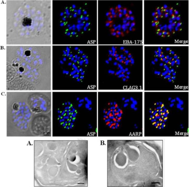
Co-localization of PfASP in late stage P. falciparum schizonts with microneme protein, EBA-175 (A); rhoptry bulb protein, Clag3.1 (B); and rhoptry neck protein, PfAARP (C). P. falciparum schizonts were stained with anti-PfASP-RIV mouse sera (green) and either anti-EBA-175 (red), anti-CLAG3.1 (red) or anti PfAARP (red) rabbit sera. Parasite nuclei were stained with DAPI (blue). PfASP did not co-localize with microneme protein EBA-175 or rhoptry bulb protein CLAG3.1 but co-localized with rhoptry neck protein PfAARP in segmented P. falciparum merozoites within late-stage schizonts. Bottom: Ultra thin sections of late-stage P. falciparum schizonts containing segmented merozoites were labelled with anti-PfASP-RIV mouse sera and gold-conjugated anti-mouse IgG. Labelling was observed in the neck of rhoptries in segmented merozoites of late-stage P. falciparum schizonts. Scale bar = 250 nm.Srivastava A, Singh S, Dhawan S, Alam MM, Mohmmed A, Chitnis CE. Localization of apical Sushi Protein in Plasmodium falciparum merozoites. Mol Biochem Parasitol. 2010 74(1):66-9 Copyright Elsevier2011
See original on MMP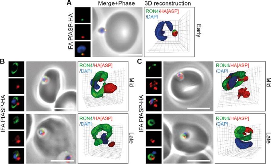
PfASP shows a dual localisation to both the tight junction and merozoite apex during erythrocyte invasion. Widefield 3D imaging of ASP-HA merozoites labeled with anti-HA, anti-PfRON4 and DAPI showing early invasion events (A) and two classes of mid/late invasion (B, C) distributions seen during invasion. Scale bar = 5 mm. 3D reconstruction with 0.2 mm grid intervals. In apically attached parasites, PfASP-HA was located at the apical tip of the merozoite at the same point as PfRON4 (A, Early), consistent with its localisation by IEM to the rhoptry neck in intracellular parasites. Mid invasion, however, when PfRON4 labeling expands to form a ring around invading parasites at the tight junction, PfASPHA was found either predominantly retained at the apical tip of the merozoite or associated with the tight junction (B, C).Zuccala ES, Gout AM, Dekiwadia C, Marapana DS, Angrisano F, Turnbull L, Riglar DT, Rogers KL, Whitchurch CB, Ralph SA, Speed TP, Baum J. Subcompartmentalisation of Proteins in the Rhoptries Correlates with Ordered Events of Erythrocyte Invasion by the Blood Stage Malaria Parasite. PLoS One. 2012;7(9):e46160.
See original on MMP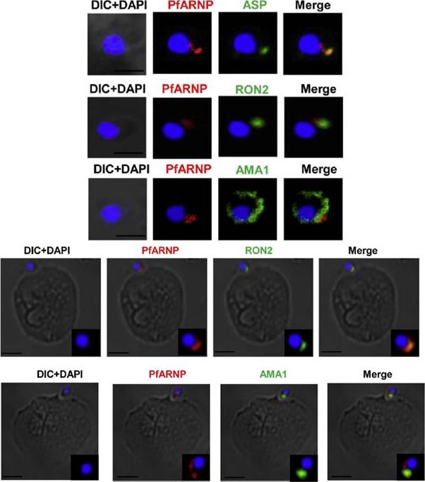
Localization of PfARNP in free merozites and at the tight junction. (Upper panel) IFA of merozoite using anti PfARNP antibody. Co-staining of PfARNP (red) was performed with ASP, RON2 (1973–2067 aa) and AMA1 (green) in free merozoites. Staining was observed at the apex of merozoite distinct from nuclear stain DAPI (blue). The staining merged perfectly well with ASP, RON2 and not with AMA1 confirming the localization of PfARNP in neck of rhoptries of merozoites. Scale bar shows 1 mm. (Lower panel) Localization of PfARNP during invasion of merozoite. IFA of PfARNP was performed with RON2 and AMA1, known markers of tight junction in Cytochalasin D treated merozoites invading to erythrocytes. The labeling of PfARNP (red) was observed at the site of attachment of merozoite to erythrocyte. The staining of PfARNP merged perfectly with RON2 and AMA1(green), indicating that PfARNP is present at the site of tight junction. The inset of each image show the optically zoomed view of invading merozoite. Scale bar shows 2 mm. Hans N, Singh S, Jain SK, Chauhan VS. Identification of novel rhoptry neck protein of Plasmodium falciparum. Mol Biochem Parasitol. 2013 188(1):34-9 Copyright Elsevier
See original on MMP
PfARNP is localized in rhoptry neck of P. falciparum. Subcellular localization of PfARNP was studied by co-staining with antibodies against merozoite surface protein-119 (A), micronemal resident protein EBA-175 (B), rhoptry bulb protein Rh2b (C), and rhoptry neck, apical sushi protein (ASP) (D). P. falciparum schizonts were co-stained with anti-PfARNP (red) and anti-MSP-119, anti- EBA-175, anti-Rh2b, anti-ASP antibodies (green). The nuclei of schizonts were stained with DAPI (blue). All apical marker proteins and PfARNP showed punctate staining in schizonts distinct from DAPI. Co-staining of PfARNP with surface marker MSP-119 showed PfARNP localized at the apical tip with surface of merozoites stained by MSP-119. PfARNP staining did not merged with either EBA-175 or Rh2b indicating that it is neither a resident of micronemes or rhoptry bulb of merozoites. The labeling of PfARNP merged perfectly with ASP confirming its localization in the neck of rhoptries of P. falciparum. (Scale bar shows 2 mm). The panel on extreme right shows digital zoom of corresponding merged images.Hans N, Singh S, Jain SK, Chauhan VS. Identification of novel rhoptry neck protein of Plasmodium falciparum. Mol Biochem Parasitol. 2013 188(1):34-9
See original on MMP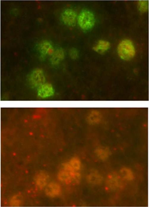
IFA experiments regarding polyclonal antibody reactivity revealed antigens expressed in mature schizonts. (E) Pre-immune serum non-reactivity.Vanegas M, Bermúdez A, Guerrero YA, Cortes-Vecino JA, Curtidor H, Patarroyo ME, Lozano JM. Protecting capacity against malaria of chemically defined tetramer forms based on the Plasmodium falciparum apical sushi protein as potential vaccine components. Biochem Biophys Res Commun. 2014 Jul 22.. [Epub ahead of print]
See original on MMPMore information
| PlasmoDB | PF3D7_0405900 |
| GeneDB | PF3D7_0405900 |
| Malaria Metabolic Pathways | Localisation images Pathways mapped to |
| Previous ID(s) | MAL4P1.58, PFD0290c, PFD0295c |
| Orthologs | PBANKA_1003600 , PCHAS_1004500 , PKNH_0304000 , PVP01_0305300 , PVX_000945 , PY17X_1005000 |
| Google Scholar | Search for all mentions of this gene |