PF3D7_0402300 reticulocyte binding protein homologue 1 (RH1)
Disruptability [+]
| Species | Disruptability | Reference | Submitter |
|---|---|---|---|
| P. falciparum 3D7 |
Possible |
15612925 | Theo Sanderson, Wellcome Trust Sanger Institute |
| P. falciparum 3D7 |
Possible |
USF piggyBac screen (Insert. mut.) | USF PiggyBac Screen |
Mutant phenotypes [+]
| Species | Stage | Phenotype | Reference | Submitter |
|---|---|---|---|---|
| P. falciparum 3D7 | Asexual |
No difference |
15612925 | Theo Sanderson, Wellcome Trust Sanger Institute |
Imaging data (from Malaria Metabolic Pathways)
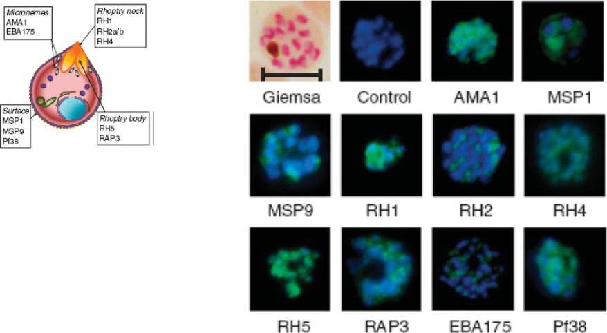
Indirect immunofluorescence images of P. falciparum schizonts stained with rabbit IgG (green) induced by 10 viral-vectored vaccines expressing malaria antigens, and negative control vectors lacking a malaria antigen. Nuclei are counterstained with 4,6-diamidino-2-phenylindole (blue). All sera were tested against 3D7 strain parasites, with the exception of anti-PfRH1 for which FVO strain parasites were used. All images to same scale as Giemsa stained image (top left, on which scale bar indicates 5 μm).Douglas AD, Williams AR, Illingworth JJ, Kamuyu G, Biswas S, Goodman AL, Wyllie DH, Crosnier C, Miura K, Wright GJ, Long CA, Osier FH, Marsh K, Turner AV, Hill AV, Draper SJ. The blood-stage malaria antigen PfRH5 is susceptible to vaccine-inducible cross-strain neutralizing antibody. Nat Commun. 2011 2:601.
See original on MMP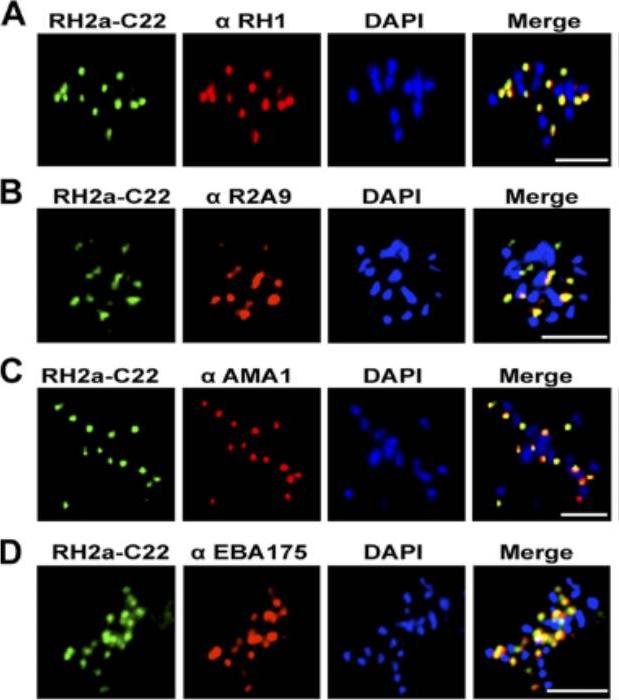
An immunofluorescence assay was performed on late-schizont-stage W2mef parasites stained for RH2a using the anti-RH2a MAb C22 and costained either with an antibody against the rhoptry neck marker RH1 (A), rabbit polyclonal antibody R2A9, recognizing both RH2a and RH2b (B), or an antibody against the micronemal marker AMA1 (C) or EBA175 (D). Bars, 5 mm. PfRH2a showed a punctate pattern at the apical tip of the merozoite in schizonts and appeared to colocalize with PfRH1 in W2mef and 3D7 parasites, strongly suggesting that RH2a is located at the rhoptry neck.Gunalan K, Gao X, Liew KJ, Preiser PR. Differences in erythrocyte receptor specificity of different parts of the Plasmodium falciparum reticulocyte binding protein homologue 2a. Infect Immun. 2011 79:3421-30.
See original on MMP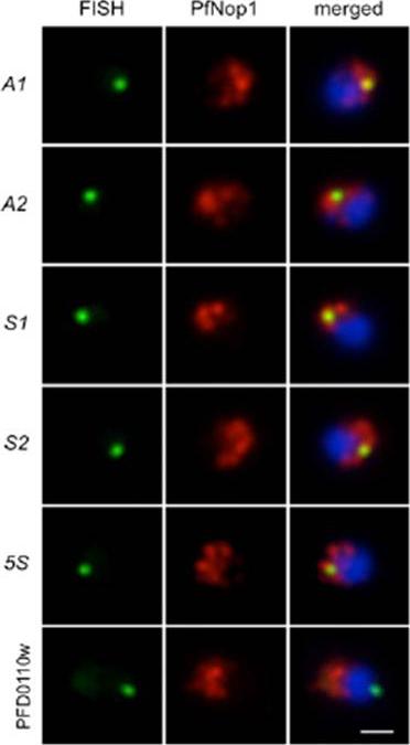
Immunofluorescence analysis using antibodies against the nucleolar protein PfNop1 (red) combined with DNA FISH (green) for individual rRNA genes (A1, A2, S1, S2, and 5S) and a subtelomeric unrelated gene (PFD0110w) on chromosome 4. The percentage of signals that localized in or near the nucleolus and the number of nuclei analyzed are as follows: A1 93.2%, n = 73; A2 90.5%, n = 95; S1 92.3%, n = 91; S2 90.0%, n = 80; 5S 90.6%, n = 128; PFD0110w 24.6%, n = 57. Parasites are in ring stage, and nuclear DNA is stained with DAPI (blue). (Scale bar, 1 μm.Mancio-Silva L, Zhang Q, Scheidig-Benatar C, Scherf A. Clustering of dispersed ribosomal DNA and its role in gene regulation and chromosome-end associations in malaria parasites. Proc Natl Acad Sci U S A. 2010 107(34):15117-22.
See original on MMP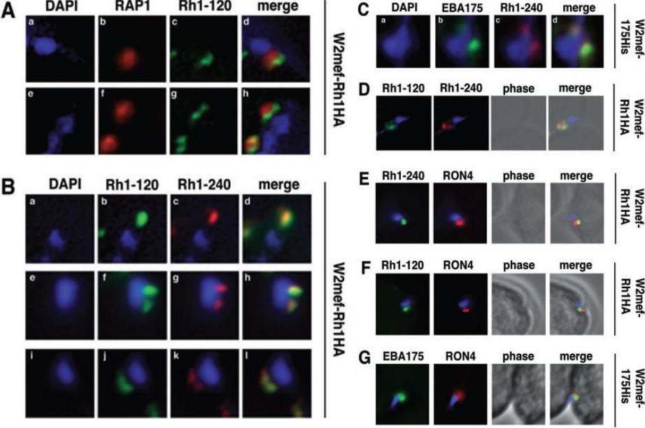
Schizont-processed Rh1-240 colocalizes with Rh1-120 at the apical tip and tight junction of the merozoite. A. The Rh1-120 protein is located at the apical tip and tight junction of merozoites. Free merozoites from the W2mef-Rh1HA line were dual stained with a McAb to RAP1 in the body of the rhoptry and with rat anti-HA antibodies to the tagged 120 kDa Rh1. Anti-HA staining is seen either apical to the RAP1 staining (top panels) or at both sides but posterior to RAP1 staining indicating tight junction staining (bottom panels). Nuclei were stained with DAPI. B. The Rh1-120 protein colocalizes with the Rh1-240 protein at the apical tip and tight junction of merozoites. Free merozoites from the W2mef-Rh1HA line were dual-stained with rat anti-HA antibodies to the tagged 120 kDa Rh1 and with a rabbit antibody (R515) to the 240 kDa Rh1. Colocalization is seen at the apical tip (panels a–d) and at the tight junction (panels e–l) of individual merozoites. W2mef-175His line C. The Rh1-240 protein colocalizes with EBA-175 MAL7P1.176 at the tight junction of merozoites. Free merozoites were dual-stained with mouse anti-His antibodies to tagged EBA-175 and with a rabbit antibody to the 240 kDa Rh1. EBA-175 and Rh1-240 appear to colocalize at the tight junction. D. Free merozoites were dual-stained with rat anti-HA antibodies to the tagged 120 kDa Rh1 and with a rabbit antibody to the 240 kDa Rh1. Simultaneous staining of both the apical tip and the tight junction of a merozoite is seen. E–G. Merozoites were trapped during invasion using cytochalasin D. Rh1-240 was stained with R515, Rh1-120 with rat anti-HA and EBA-175 with mouse anti-His antibodies. PfRON4 was stained either with a mouse antibody (E) or with a rabbit antibody (F and G). Triglia T, Tham WH, Hodder A, Cowman AF. Reticulocyte binding protein homologues are key adhesins during erythrocyte invasion by Plasmodium falciparum. Cell Microbiol. 2009 11:1671-87. Copyright John Wiley & Sons Ltd. 2010.
See original on MMP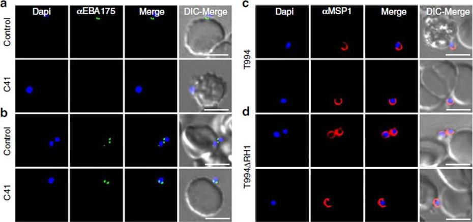
Effect of anti-PfRH1 inhibitory mAbs on merozoite surface expression of EBA175 during invasion. (a–d) Surface expression of EBA175 and MSP1 on T994 and T994DRH1 was detected by immunofluorescence assays. Isolated T994 (a,c) or T994DRH1 (b,d) merozoites were directly incubated with erythrocytes containing 4 mM Cyto D in the absence (Control) and presence (C41) of C41 (0.2mgml1) before analysis of expression of EBA175 and MSP1 at the junction. Both differential interference contrast (DIC) and fluorescence images were captured. Nuclear DNA was counterstained with DAPI (blue). EBA175 is shown in green, whereas MSP1 is shown in red. Fluorescence images, merged fluorescence images (DAPI and green or DAPI with red) and merged fluorescence images with DIC images are shown. Scale bars=10 mM. Antibody C41 blocks surface expression of EBA175 in T994 (a lower panel), whereas it has no effect on T994DRH1 parasites (b lower panel). In the absence of C41, EBA175 is expressed on the surface of both T994 and T994DRH1 (upper panels a,b). Ab has no effect on the detection of MSP1 on the surface of merozoites (c,d).Gao X, Gunalan K, Yap SS, Preiser PR. Triggers of key calcium signals during erythrocyte invasion by Plasmodium falciparum. Nat Commun. 2013;4:2862.
See original on MMP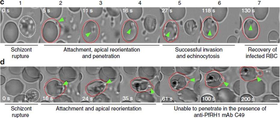
Anti-PfRH1 mAbs inhibit invasion before junction formation. (c,d) Snapshots taken from time-lapse live movie microscopy of invasion of W2mef by merozoites in the absence (c) or presence (d) of C49 mAb. Time-elapsed post schizont rupture is indicated in each snapshot in seconds (sec, white). A white dotted circle indicates a rupturing schizont. Green arrows point to merozoites. A red dotted circle around an erythrocyte is added to help follow the infection. Scale bars¼5 mM. (c) In the absence of mAb, the merozoite released from a mature schizont (white cycle) attaches, apically reorients and penetrates into an uninfected erythrocyte. Following successful invasion, deformation of infected erythrocytes occurs (echinocytosis) and by 2 min the erythrocytes recover back to its normal shape. (d) In the presence of C49, the merozoite released from a mature schizont attaches and apically reorients, but it is unable to penetrate the erythrocyte even after 3 min.Gao X, Gunalan K, Yap SS, Preiser PR. Triggers of key calcium signals during erythrocyte invasion by Plasmodium falciparum. Nat Commun. 2013;4:2862.
See original on MMP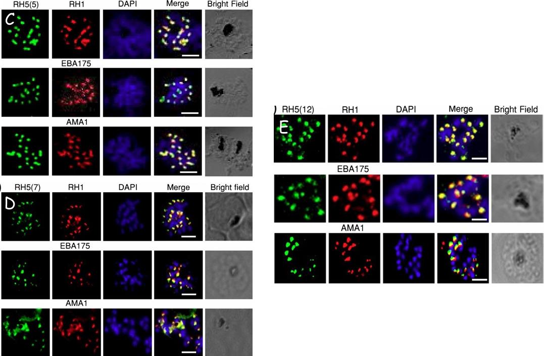
Validation of RH5 mAbs by Immunofluroscence Microscopy. Smears of 3D7 late stage schizont were co- stained with RH5(5) (c) (7) (d) and (12) (e) (first column, green) and antibodies against other invasion markers RH1, EBA175 and AMA1 respectively (second column, red). Parasite nuclei were stained with DAPI (blue). In the merged images, areas of overlap between the red and the green signals are shown in yellow. Bright field indicates the outline of the schizont. Scale bar =2μm.Aniweh Y, Gao X, Hao P, Meng W, Lai SK, Gunalan K, Chu TT, Sinha A, Lescar J, Chandramohanadas R, Li HY, Sze SK, Preiser PR. RH5-Basigin interaction induceschanges in the cytoskeleton of the host RBC. Cell Microbiol. 2017 Apr 13 [Epub ahead of print] PMID: 28409866
See original on MMPMore information
| PlasmoDB | PF3D7_0402300 |
| GeneDB | PF3D7_0402300 |
| Malaria Metabolic Pathways | Localisation images Pathways mapped to |
| Previous ID(s) | MAL4P1.22, PFD0110w |
| Orthologs | |
| Google Scholar | Search for all mentions of this gene |