PF3D7_0402200 surface-associated interspersed protein 4.1 (SURFIN 4.1), pseudogene (SURF4.1)
Disruptability [+]
| Species | Disruptability | Reference | Submitter |
|---|---|---|---|
| P. falciparum 3D7 |
Possible |
USF piggyBac screen (Insert. mut.) | USF PiggyBac Screen |
Mutant phenotypes [+]
| Species | Stage | Phenotype | Reference | Submitter |
|---|---|---|---|---|
| P. falciparum 3D7 | Asexual |
No difference |
28800640 (Knock down)
Initially increased transcript abundance |
Theo Sanderson, Wellcome Trust Sanger Institute |
| P. falciparum 3D7 | Asexual |
Invasion defect |
https://www.biorxiv.org/content/biorxiv/early/2019/02/27/562124.full.pdf
(Knock down)
"Transcript knockdown revealed that surf4.1 20 is essential for merozoite formation in late trophozoite/schizont stages while DNA replication 21 seemed not to be influenced" |
Theo Sanderson, Google AI |
| P. falciparum 3D7 | Asexual |
Refractory |
https://www.biorxiv.org/content/biorxiv/early/2019/02/27/562124.full.pdf
(Insert. mut.)
Attempts to introduce an additional stop codon into this pseudogene were unsuccessful |
Theo Sanderson, Google AI |
Imaging data (from Malaria Metabolic Pathways)
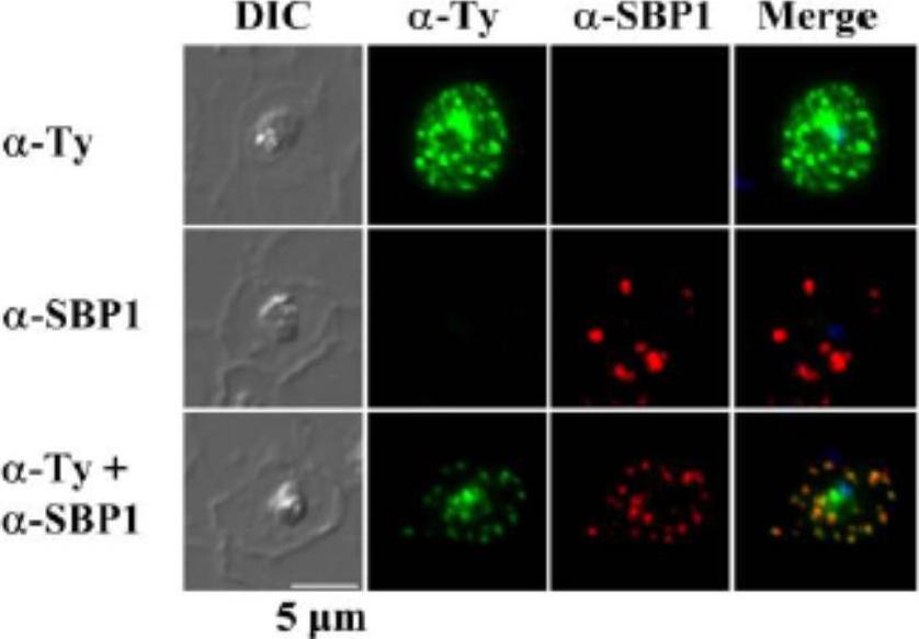
Subcellular localization of rSURFIN4.1 Full (3D7A) in P. falciparum-infected red blood cells. Representative fluorescence images showing the localization of rSURFIN4.1 The differential interference contrast (DIC), fluorescence image with mouse anti-Ty antibody (α-Ty, reacts with Surfin4.1), Maurer’s clefts location with rabbit anti-SBP1 antibody (α-SBP1), and merged image with nucleus staining with DAPI (Merge) are shown. rSURFIN4.1 exhibits a dotted fluorescence pattern within the red blood cell cytosol, co-localized with PfSBP1. Bar = 5 μm.Zhu X, Yahata K, Alexandre JS, Tsuboi T, Kaneko O. The N-terminal segment of Plasmodium falciparum SURFIN(4.1) is required for its trafficking to the red blood cell cytosol through the endoplasmic reticulum. Parasitol Int. 2013 62(2):215-29
See original on MMP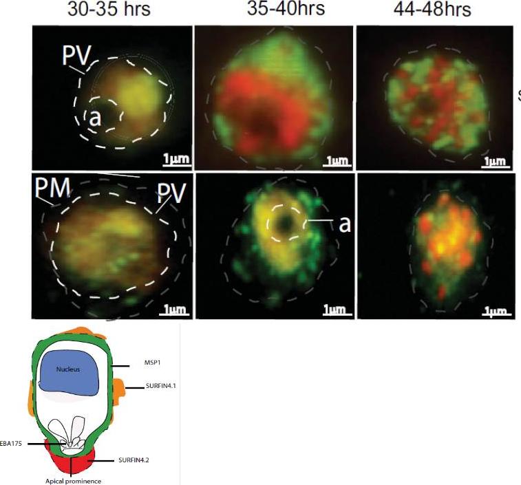
Localization of SURFIN4.1 by immunofluorescence staining on air-dried monolayers from 3D7S8 parasite strain. Air dried monolayers probed with rabbit anti-SURFIN4.1 on 3D7S8 pRBC. Propidium iodide (red) was used to stain the parasite nucleus and SURFIN4.1 and SURFIN4.2 (which is encoded by the same gene) proteins were stained green using anti-rabbit Alexa 488. SURFIN4.1 localizes within the parasitophorous vacuole (PV) and is observed from approximately 30 hrs post invasion. SURFIN4.1 was observed as a green dot above the food vacuole (a), at 30–35 hrs parasite stages. The protein was spread around the parasitophorous vacuole (PV) at 35–40 hrs parasite stages and in the mature schizont (44–48 hrs) SURFIN4.1 was observed between the dividing merozoites. During the trophozoite and early schizont stages SURFIN4.2 shows a similar pattern of staining as SURFIN4.1.Mphande FA, Ribacke U, Kaneko O, Kironde F, Winter G, Wahlgren M. SURFIN4.1, a schizont-merozoite associated protein in the SURFIN family of Plasmodium falciparum. Malar J. 2008 Jul 1;7:116. PMID:
See original on MMP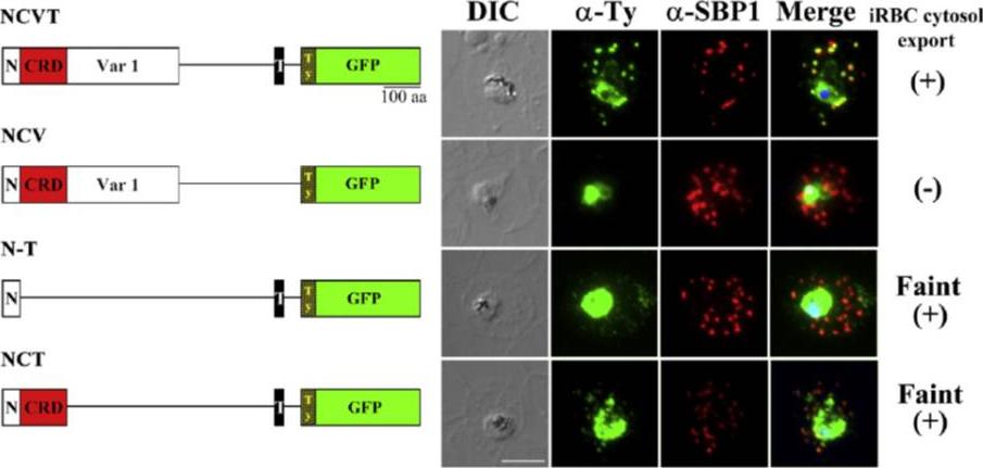
Regions of SURFIN4.1 required for the export to the Maurer's clefts. (A) Schematic drawings of the recombinant proteins, rSURFIN4.1 NCVT (NCVT), rSURFIN4.1 NCV (NCV), rSURFIN4.1 NCT (NCT), and rSURFIN4.1 N-T (N-T) are depicted in the left panels. The right panels show co-localization of a series of recombinant SURFIN4.1 detected with anti-Ty antibody (α-Ty) and counter-staining with PfSBP1 (α-SBP1) by IFA. The differential interference contrast (DIC) and merged images with nucleus stained with DAPI (Merge) are shown. (+) or (−) indicates positive or negative detection of the signals in the iRBC cytosol, respectively. Bar=5 μm. dotted fluorescence signal pattern in the iRBC cytosol, which co-localized with a MC marker PfSBP1. TM region is essential for export from the parasite to the PV lumenZhu X, Yahata K, Alexandre JS, Tsuboi T, Kaneko O. The N-terminal segment of Plasmodium falciparum SURFIN4.1 is required for its trafficking to the red blood cell cytosol through the endoplasmic reticulum. Parasitol Int. 2012 62(2):215-229.
See original on MMP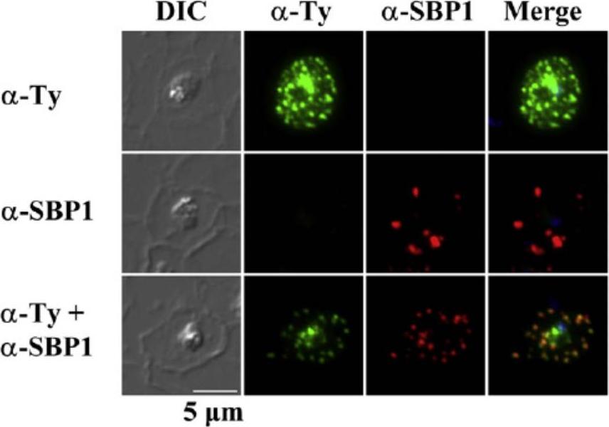
Subcellular localization of rSURFIN4.1Full (3D7A) in P. falciparum-infected red blood cells. Representative fluorescence images showing the localization of rSURFIN4.1Full. The differential interference contrast (DIC), fluorescence image with mouse anti-Ty antibody (α-Ty; recognizing rSURFIN4.1), Maurer's clefts location with rabbit anti-SBP1 antibody (α-SBP1), and merged image with nucleus staining with DAPI (Merge) are shown. rSURFIN4.1Full exhibits a dotted fluorescence pattern within the red blood cell cytosol, co-localized with PfSBP1. Bar=5 μm.Zhu X, Yahata K, Alexandre JS, Tsuboi T, Kaneko O. The N-terminal segment of Plasmodium falciparum SURFIN4.1 is required for its trafficking to the red blood cell cytosol through the endoplasmic reticulum. Parasitol Int. 2012 62(2):215-229.
See original on MMPMore information
| PlasmoDB | PF3D7_0402200 |
| GeneDB | PF3D7_0402200 |
| Malaria Metabolic Pathways | Localisation images Pathways mapped to |
| Previous ID(s) | 3D7Surf4.1, MAL4P1.20, MAL4P1.21, PFD0100c, PFD0105c |
| Orthologs | |
| Google Scholar | Search for all mentions of this gene |