PF3D7_0309600 60S acidic ribosomal protein P2 (PfP2)
Disruptability [+]
| Species | Disruptability | Reference | Submitter | |
|---|---|---|---|---|
| P. falciparum 3D7 |
Refractory |
USF piggyBac screen (Insert. mut.) | USF PiggyBac Screen | |
| P. berghei ANKA |
Refractory |
RMgm-768 | Imported from RMgmDB | |
| P. berghei ANKA |
Refractory |
PlasmoGEM (Barseq) | PlasmoGEM | |
Mutant phenotypes [+]
None reported yet. Please press the '+' button above to add one.Imaging data (from Malaria Metabolic Pathways)
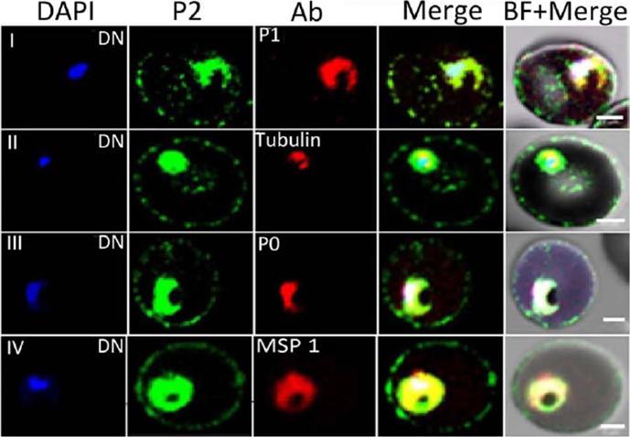
Immunofluorescence assay of P. falciparum infected erythrocytes (IE) using various antibodies. Localization of DAPI (blue), P2 (green); various antibodies (anti-PfP1, anti b-tubulin, anti-PfP0, and anti-MSP1) in red at the di-nuclear (DN) stage of Plasmodium falciparum infected erythrocytes. Scale bar indicates 2 mm.Das S, Basu H, Korde R, Tewari R, Sharma S. Arrest of Nuclear Division in Plasmodium through Blockage of Erythrocyte Surface Exposed Ribosomal Protein P2. PLoS Pathog. 2012 Aug;8(8):e1002858.
See original on MMP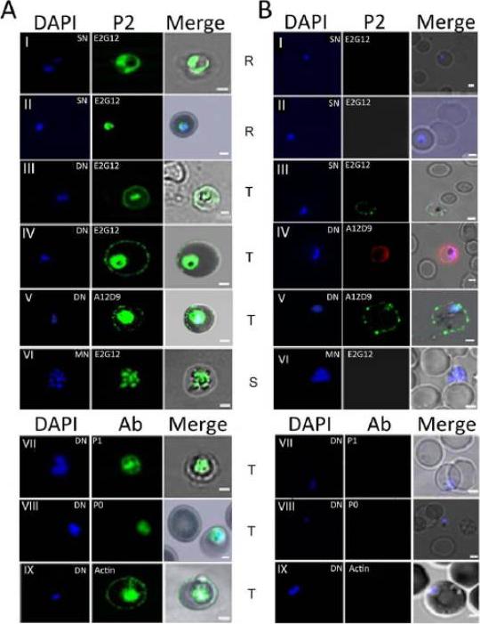
Immunofluorescence assay of P. falciparum infected erythrocytes (IE) using various antibodies. (A) & (B) show confocal images of permiabilized (IFA) and unpermeabilized in solution (SIFA) assays respectively, of different IE stages (I, II: Ring; III, IV, V, VII, VIII, IX: trophozoite; VI: schizont) IFA analysis of permeabilized IEs from asynchronous cultures of P. falciparum 3D7 showed a transient presence of P2 protein on the IE-surface at certain stages of development (A). This was often coincident with a dumbbell shape, elongated or crescent shaped DAPI stained nuclei (henceforth designated as the di-nuclear or DN-stage). It was not seen with the tightly packed single nuclear (SN) rings, nor the multi-nuclear (MN) schizont stage IE-surfaces (A). Cytoplasmic P2 staining was observed at all stages (A). To test for surface exposure, unpermeabilized solution immunofluoroscence assay (SIFA) was carried out which confirmed the presence of P2 on the outer IE surface at the DN stage but not at the SN or MN stages (B). Two different anti-P2 mAbs, E2G12 and A12D9, as well as two different secondary antibodies with different flurophores (Alexa 488 and 594) showed similar results, confirming the specific surface reactivity of PfP2 protein.Das S, Basu H, Korde R, Tewari R, Sharma S. Arrest of Nuclear Division in Plasmodium through Blockage of Erythrocyte Surface Exposed Ribosomal Protein P2. PLoS Pathog. 2012 Aug;8(8):e1002858.
See original on MMP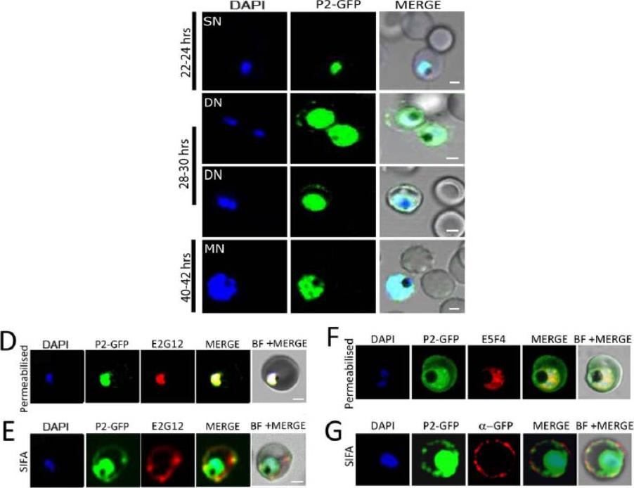
Upper panel: IFA of synchronized P. falciparum cells using DAPI (blue), P2 (green), and bright field of IE at various time points PMI in parasite development. Scale bar indicates 2 mm. Confocal microscopy of P2-GFP transfected cells at different stages of parasite growth. DAPI (blue), P2-GFP (green). GFP on the IE-surface at 28–30 hrs PMI trophozoite stage, but not at 22–24 hrs or 40–42 hrs.Lower panel: (D,F) Confocal microscopy of permeabilized IE showing D) Red: anti-PfP2 mAb E2G12; F) Red: anti-PfP0 mAb E5F4; Green: P2-GFP staining of Pf3D7 P2-GFP infected RBCs. (E,G) Solution immunofluorescence (SIFA) of DN Pf3D7 P2-GFP infected RBCs showing E) Red: anti-PfP2 mAb E2G12; G) Red: anti-GFP antibody; Green: P2-GFP staining of Pf3D7 P2-GFP infected RBCs. Unpermeabilized SIFA at the DN stage showed that the P2 staining and GFP colocalized on the IE surface (4E).Das S, Basu H, Korde R, Tewari R, Sharma S. Arrest of Nuclear Division in Plasmodium through Blockage of Erythrocyte Surface Exposed Ribosomal Protein P2. PLoS Pathog. 2012 Aug;8(8):e1002858.
See original on MMP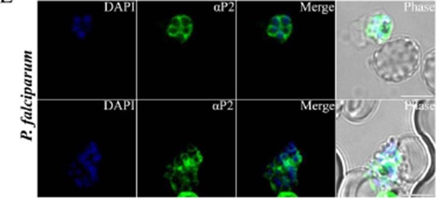
P. falciparum schizonts stained with αP2 mouse polyclonal antibodies. Nuclei stained with DAPI. Scale bars represent 5 μm. Distribution of P2 in schizonts at the periphery of themerozoites, giving a ‘honey-comb’ like appearance,which is characteristic of merozoite surface proteins. Thus it appeared that in addition to the presence of PfP2 at the infected erythrocyte surface at the onset of cell division, PfP2 was also present on the merozoite surface. Sudarsan R, Chopra RK, Khan MA, Sharma S. Ribosomal protein P2 localizes to the parasite zoite-surface and is a target for invasion inhibitory antibodies in Toxoplasma gondii and Plasmodium falciparum. Parasitol Int. 2014 Sep 30 [Epub ahead of print]
See original on MMPMore information
| PlasmoDB | PF3D7_0309600 |
| GeneDB | PF3D7_0309600 |
| Malaria Metabolic Pathways | Localisation images Pathways mapped to |
| Previous ID(s) | MAL3P3.19, PFC0400w |
| Orthologs | PBANKA_0407700 , PCHAS_0408600 , PKNH_0833100 , PVP01_0830600 , PVX_119587 , PY17X_0410200 |
| Google Scholar | Search for all mentions of this gene |